

Pillars article: the CD4 receptor is complexed in detergent lysates to a protein-tyrosine kinase (pp58) from human T lymphocytes. 1988
- PMID: 20724730
- PMCID: PMC3791413
Pillars article: the CD4 receptor is complexed in detergent lysates to a protein-tyrosine kinase (pp58) from human T lymphocytes. 1988
Abstract
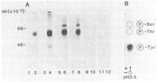
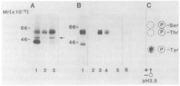
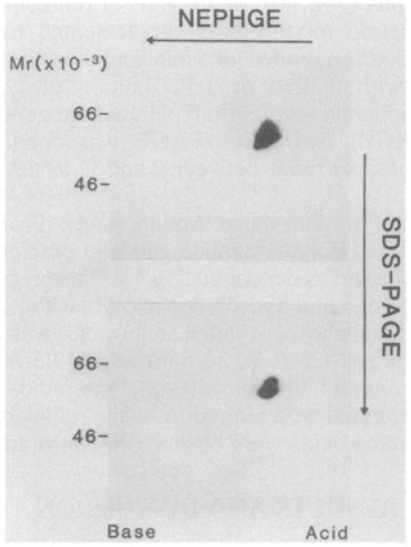
The CD4 (T4) antigen is a cell-surface glycoprotein that is expressed predominantly on the surface of helper T cells and has been implicated in the regulation of T-cell activation and in the associative recognition of class II antigens of the major histocompatibility complex. In addition, the CD4 antigen appears to serve as a receptor for the human immunodeficiency virus (HIV). An important question has been whether the CD4 receptor is linked to an intracellular mediator that could regulate the activation of the CD4+ subset. In this paper, we provide preliminary evidence that the CD4 receptor is complexed in detergent lysates to a protein-tyrosine kinase (PTK) of 55–60 kDa, which is expressed specifically in T cells. The PTK is the human analogue of the murine pp56LSTRA (pp56lck) and has significant homology with c-src, c-yes, and other members of the src family. The identification of the PTK associated with CD4 receptor was made by use of an antiserum to a synthetic peptide that was deduced from the DNA sequence of PTK. Two-dimensional nonequilibrium pH gradient gel electrophoresis/NaDodSO4/PAGE revealed the kinase to focus as a heterogeneous collection of spots in the pH range of 4.0–5.0. Furthermore, in vitro phosphorylation revealed the phosphorylation of two additional polypeptides at 40 and 80 kDa, in addition to the autophosphorylation of the PTK at 55–60 kDa. The potential importance of the association between the CD4 receptor and the PTK of T cells is discussed in relation to T-cell activation and HIV infectivity.
Figures
Comment in
-
Multiple roles of CD4 and CD8 in T cell activation.J Immunol. 2010 Sep 1;185(5):2643-4. doi: 10.4049/jimmunol.1090076. J Immunol. 2010. PMID: 20724729 No abstract available.
Similar articles
-
The CD4 receptor is complexed in detergent lysates to a protein-tyrosine kinase (pp58) from human T lymphocytes.Proc Natl Acad Sci U S A. 1988 Jul;85(14):5190-4. doi: 10.1073/pnas.85.14.5190. Proc Natl Acad Sci U S A. 1988. PMID: 2455897 Free PMC article.
-
Pillars article: the CD4 and CD8 T cell surface antigens are associated with the internal membrane tyrosine-protein kinase p56lck. 1994.J Immunol. 2010 Sep 1;185(5):2650-7. J Immunol. 2010. PMID: 20724731 No abstract available.
-
An octapeptide analogue of HIV gp120 modulates protein tyrosine kinase activity in activated peripheral blood T lymphocytes.Clin Exp Immunol. 1995 Jun;100(3):412-8. doi: 10.1111/j.1365-2249.1995.tb03715.x. Clin Exp Immunol. 1995. PMID: 7539724 Free PMC article.
-
HIV1 Nef: the Machiavelli of cellular activation.Res Virol. 1997 Jan-Feb;148(1):58-64. doi: 10.1016/s0923-2516(97)81915-2. Res Virol. 1997. PMID: 9017836 Review. No abstract available.
-
Regulation of Nef function.Res Virol. 1997 Jan-Feb;148(1):64-8. doi: 10.1016/s0923-2516(97)81916-4. Res Virol. 1997. PMID: 9017837 Review. No abstract available.
Cited by
-
Dynamics of the Coreceptor-LCK Interactions during T Cell Development Shape the Self-Reactivity of Peripheral CD4 and CD8 T Cells.Cell Rep. 2020 Feb 4;30(5):1504-1514.e7. doi: 10.1016/j.celrep.2020.01.008. Cell Rep. 2020. PMID: 32023465 Free PMC article.
-
LFA-1 activates focal adhesion kinases FAK1/PYK2 to generate LAT-GRB2-SKAP1 complexes that terminate T-cell conjugate formation.Nat Commun. 2017 Jul 12;8:16001. doi: 10.1038/ncomms16001. Nat Commun. 2017. PMID: 28699640 Free PMC article.
-
How the Discovery of the CD4/CD8-p56lck Complexes Changed Immunology and Immunotherapy.Front Cell Dev Biol. 2021 Mar 15;9:626095. doi: 10.3389/fcell.2021.626095. eCollection 2021. Front Cell Dev Biol. 2021. PMID: 33791292 Free PMC article. Review.
-
The Immune Adaptor SLP-76 Binds to SUMO-RANGAP1 at Nuclear Pore Complex Filaments to Regulate Nuclear Import of Transcription Factors in T Cells.Mol Cell. 2015 Sep 3;59(5):840-9. doi: 10.1016/j.molcel.2015.07.015. Epub 2015 Aug 27. Mol Cell. 2015. PMID: 26321253 Free PMC article.
References
-
- Reinherz EL, Meuer SC, Schlossman SF. Immunol. Rev. 1983;74:83–112. - PubMed
-
- Swain SL. Immunol. Rev. 1983;74:129–142. - PubMed
-
- Dalgleish A, Beverley PCL, Clapham PR, Crawford DH, Greaves MF, Weiss RA. Nature (London) 1984;312:763–766. - PubMed
-
- Klatzman D, Champayne E, Chamaret S, Gruest J, Guefard D, Hercend T, Gluckman JC, Montagnier L. Nature (London) 1984;312:767–768. - PubMed
-
- Maddon P, Dalgleish AG, McDougal JS, Clapham PR, Weiss RA, Axel R. Cell. 1986;47:333–348. - PubMed
Publication types
MeSH terms
Substances
Personal name as subject
- Actions
- Actions
- Actions
- Actions
- Actions
Grants and funding
LinkOut - more resources
Full Text Sources
Research Materials
Miscellaneous