. 2010 Oct;6(7):891-900.
doi: 10.4161/auto.6.7.13038. Epub 2010 Oct 16.

Autophagic degradation of active caspase-8: a crosstalk mechanism between autophagy and apoptosis

Affiliations

Autophagic degradation of active caspase-8: a crosstalk mechanism between autophagy and apoptosis

Wen Hou et al. Autophagy. 2010 Oct.

Abstract

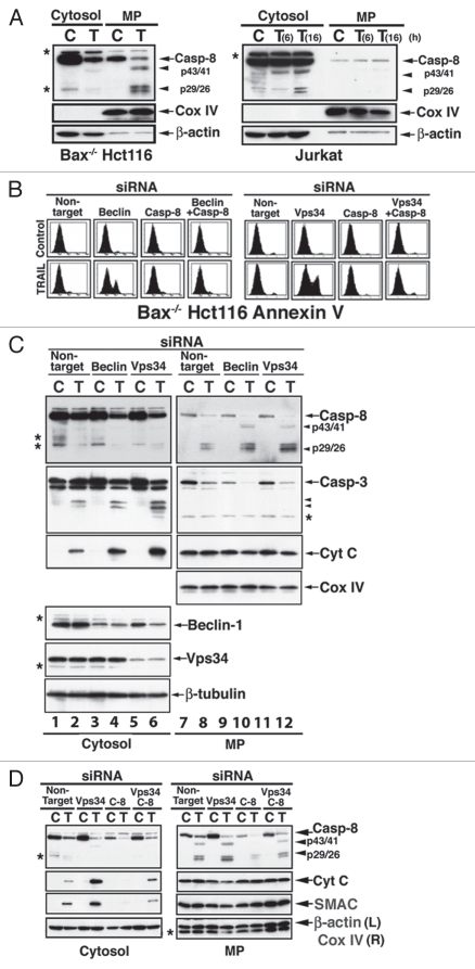
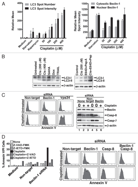
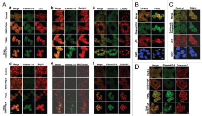
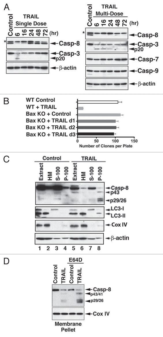
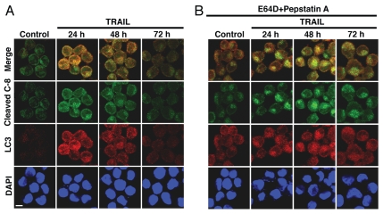
Apoptotic defects endow tumor cells with survival advantages. Such defects allow the cellular stress response to take the path of cytoprotective autophagy, which either precedes or effectively blocks an apoptotic cascade. Inhibition of the cytoprotective autophagic response shifts the cells toward apoptosis, by interfering with an underlying molecular mechanism of cytoprotection. The current study has identified such a mechanism that is centered on the regulation of caspase-8 activity. The study took advantage of Bax(-/-) Hct116 cells that are TRAIL-resistant despite significant DISC processing of caspase-8, and of the availability of a caspase-8-specific antibody that exclusively detects the caspase-8 large subunit or its processed precursor. Utilizing these biological tools, we investigated the expression pattern and subcellular localization of active caspase-8 in TRAIL-mediated autophagy and in the autophagy-to-apoptosis shift upon autophagy inhibition. Our results suggest that the TRAIL-mediated autophagic response counter-balances the TRAIL-mediated apoptotic response by the continuous sequestration of the large caspase-8 subunit in autophagosomes and its subsequent elimination in lysosomes. The current findings are the first to provide evidence for regulation of caspase activity by autophagy and thus broaden the molecular basis for the observed polarization between autophagy and apoptosis.


Figures

Figure 1
Figure 1
Caspase-8 requirement for the shift from TRAIL-mediated autophagy to TRAIL-mediated apoptosis. (A) Cell-specific subcellular localization of cleaved caspase-8. Processed fragments of cleaved caspase-8 are associated with a membranous pellet (MP) in TRAIL-treated Bax-/- Hct16 cells (100 ng/ml 6 hr, left part, C = Control; T = TRAIL), but with cytosol in TRAIL-treated Jurkat cells (25 ng/ml, right part). Mitochondrial inner membrane cytochrome c oxidase (Cox) IV and β-actin served as loading controls for their respective subcellular fractions. (B) Inhibition of TRAIL-mediated autophagy induces caspase-8-dependent apoptosis. An autophagy-to-apoptosis shift is induced by Beclin 1 KD (left part) or Vps34 KD (right part), and it is inhibited by concomitant KD of caspase-8. Bax-/- Hct116 cells were treated with the indicated siRNA for 60 hr and then with TRAIL for 6 hr. The cells were assessed by Annexin V flow cytometry. (C) Inhibition of autophagy in TRAIL-treated Bax-/- Hct116 cells is associated with increased processing of caspase-8 and caspase-3, and cytochrome c release. Bax-/- Hct116 cells were treated with the indicated siRNAs and TRAIL as described in (B), and following cytosol and MP separation, the subcellular fractions were assessed by immunoblotting for the expression of the indicated proteins. Please note, the doublet protein bands detected by anti-caspase-8 Ab in control cells (lanes 1 and 3, marked by asterisks) are unidentified, as they do not align with the doublet detected in TRAIL-treated cells (lanes 8, 10 and 12). (D) The release of cytochrome c and SMAC that is induced by a combined treatment of Vps34 siRNA and TRAIL is inhibited by caspase-8 siRNA. Experimental details are described in (A and B).
Figure 2
Figure 2
Sensitization of apoptotic-deficient MCF7 cells to cisplatin by autophagy inhibition is caspase-8-dependent. (A) Quantitation by image cytometry of drug-mediated changes in LC3 (left part) and Beclin 1 (right part) in apoptotic-deficient MCF7 cells. Cisplatin-selected MCF7 cells were treated as indicated for 12 hrs (rapamycin, 2 µM). The cells were then fixed and stained with LC3- and Beclin 1-specific Abs, and fluorescence quantitation was performed by the Cellomics ArrayScan Image Cytometer. The data are means ± SEM of quadruplet determinations of relative spot intensity or spot number per 500 cells counted in each well. Similar results were obtained in at least three independent experiments. (B) Immunoblot analysis for the presence of LC3-II in cisplatin-resistant MCF7 cells treated with cytotoxic drugs. Left part shows induced expression of LC3-II in cisplatin-selected MCF-7 freshly treated with cisplatin, SAHA (2 µM), doxorubicin (100 ng/ml); or TRAIL (100 ng/ml). Right part demonstrates that the cisplatin-mediated increase in LC3-II represents autophagic flux. Cisplatin-selected MCF7 cells were freshly treated with cisplatin (200 µM) in the presence or absence of the cathepsin inhibitors, E64D and pepstatin A. MBL anti-LC3 Ab was used for probing, and β-actin served as a loading control. (C) Inhibition of autophagy by Beclin 1 or Vps34 RNAi reverses the resistance of MCF7 cells to cisplatin. Cisplatin-resistant MCF7 cells were treated with nontarget, Beclin 1 or Vps34 siRNAs for 72 hr and then with cisplatin (200 µM) for 12 hr. The apoptotic response was assessed by Annexin V (left part) and by the level of caspase-8 processing (right part). (D) The cisplatin-sensitization by Beclin 1 siRNA is inhibited in the presence of pharmacological inhibitors of caspases (left part) or by caspase-8 RNAi (right part). The experiments were performed as described in C, and a fresh cisplatin dose was added in the presence of Z-VAD-FMK or A-IETD-FMK (100 µM each). Apoptosis was determined by Annexin V/PI flow cytometry, and one experiment of at least three with similar results is shown. In the right part, cisplatin-selected MCF7 cells were treated as described in (C) with the addition of caspase-8 siRNA. The mild increase in cell death in cells treated only with caspase-8 siRNA may relate to interference with the survival activity of caspase-8. Similar results were obtained with siGENOME SMARTpool mixture of four non-overlapping siRNAs from Dharmacon and with three additional independent caspase-8 siRNAs from Invitrogen.
Figure 3
Figure 3
The co-localization of cleaved caspase-8 subunit with components of the autophagic process increases in the presence of E64D and pepstatin A. (A) Accumulation of cleaved caspase-8 in Bax-/- Hct116 cells treated with TRAIL (100 ng/ml, 5 hr) in the presence of E64D/pepstatin A and its co-localization with LC3 (autophagosome), Beclin 1 (autophagosomal nucleation), LAMP2 (lysosome) and Bap31 (ER), but at a reduced level with MitoTracker or β-tubulin. (B) Co-localization of cleaved caspase-8 with LC3 in the minor fraction of non-apoptotic TRAIL-treated WT Hct116 cells. Cells with fragmented nuclei (major fraction) demonstrate an increase in cleaved caspase-8, but no increase in LC3. Nuclei were stained by DAPI. (C) Partial co-localization of full-length caspase-8 (N-terminus-specific mAb) with LC3 in control or TRAIL-treated Bax-/- Hct116 cells. (D) Differential localization of cleaved caspase-8 and full-length caspase-7 in TRAIL treated Hct116 Bax-/- cells. Scale bars = 40 µm. Please note that in the presence of E64D/PepA, there is an increased distinction between the localization of accumulated cleaved caspase-8 and full-length caspase-7.
Figure 4
Figure 4
The clonogenic capability of Bax-/- Hct116 cells is not affected by repeated TRAIL treatments despite continuous processing of caspase-8. (A) processing of caspase-8 in response to a single (left part) or multiple (right part) TRAIL treatments. Bax-/- Hct116 cells were treated once or daily for three days with TRAIL (100 ng/ml). Expression of the various caspases was assessed by immunoblotting. Please note, caspase-3 is processed only into p20, which remains inhibited in the absence of a mitochondrial contribution. The asterisks indicate unidentified protein bands. (B) Clonogenicity of Bax-/- Hct116 cells successively treated with a daily fresh dose of TRAIL (100 ng/ml) for 3 days as compared to WT Hct116 cells treated once with TRAIL (25 ng/ml, 6 hr). Colonies that developed during a 14-day culture in a methylcellulose-based medium in 35-mm plates were counted using an inverted microscope and gridded screen. The data (means ± SE M) were derived from three replicates in one of three experiments with equivalent results. Clonogenic assays were performed 24 hr after the first (d1), second (d2) and third (d3) TRAIL treatments of Bax-/- Hct116 cells. (C) Co-localization of cleaved caspase-8 with P-100 LC3-II during TRAIL-mediated autophagy. Control and TRAIL-treated Bax-/- Hct116 cells were subcellular fractionated to obtain purified mitochondria, S-100 and P-100 fractions. S-100 is regarded as cytosolic fraction, and P-100 as light membrane fraction. While LC3-I was detected in the S-100, LC3-II and cleaved caspase-8 were detected in the P-100 fraction. The loading in this experiment was cell-related, as indicated by the expression levels of Cox IV and β-actin. The presence of β-actin in the P-100 fraction may relate to its reported association with autophagosomes, and it serves to demonstrate an equal loading between control P-100 (lane 4) and TRAIL-treated cell P-100 (lane 8). (D) Accumulation of processed caspase-8 fragments in MP fraction of Bax-/- Hct116 cells treated with TRAIL in the presence of E64D.
Figure 5
Figure 5
Enhanced co-localization of cleaved caspase-8 with LC3-positive punctates in cells treated with TRAIL and cathepsin inhibitors. Bax-/- Hct116 cells were treated daily with TRAIL (A) or with TRAIL and E64D/pepstatin A (B). Cytospins that were prepared at the indicated time points were all stained and assessed by confocal microscopy at the same time. The co-accumulation of cleaved caspase-8 with LC3 is appreciably increased early after TRAIL treatment (24 hr) with a marked reduction in the following days (A); the accumulation of cleaved caspase-8 is most pronounced with less reduction in subsequent days in cells treated with TRAIL and E64D/pepstatin A (B).
Figure 6
Figure 6
Differential patterns of lysosomal accumulation of cleaved caspase-8 as compared to cleaved caspase-3 in Bax-/- Hct116 cells treated with TRAIL and cathepsin inhibitors. Bax-/- Hct116 cells were treated daily with a fresh dose of TRAIL (100 ng/ml) for three consecutive days in the absence (A and C) or presence (B and D) of E64D/pepstatin A. The staining was performed on adherent cells in chamber slides. Of note, degradation of cleaved caspase-8 (A and B), but not of caspase-3 (C and D), is inhibited by E64D/pepstatin A. Scale bars = 40 µm. (E and F) Quantitation of co-localization with LAMP2 of cleaved caspase-8 (E) and cleaved caspase-3 (F). The co-localization was measured by Metamorph (Molecular Devices, Downingtown, PA) and performed on cells obtained from at least three independent experiments. The data are means ± SE M of 20–25 individually analyzed stained cells per sample.

Similar articles

Cited by

References

    1. Salvesen GS, Riedl SJ. Caspase mechanisms. Adv Exp Med Biol. 2008;615:13–23. - PubMed
    1. Riedl SJ, Shi Y. Molecular mechanisms of caspase regulation during apoptosis. Nat Rev Mol Cell Biol. 2004;5:897–907. - PubMed
    1. Salvesen GS, Duckett CS. IAP proteins: blocking the road to death's door. Nat Rev Mol Cell Biol. 2002;3:401–410. - PubMed
    1. Srinivasula SM, Ashwell JD. IAPs: what's in a name? Mol Cell. 2008;30:123–135. - PMC - PubMed
    1. Cecconi F, Levine B. The role of autophagy in mammalian development: cell makeover rather than cell death. Dev Cell. 2008;15:344–357. - PMC - PubMed

Publication types

MeSH terms