

Cancer-associated retinopathy in neuroendocrine carcinoma of the fallopian tube
- PMID: 20724944
- PMCID: PMC3491900
- DOI: 10.1097/WNO.0b013e3181e22ef0
Cancer-associated retinopathy in neuroendocrine carcinoma of the fallopian tube
Abstract
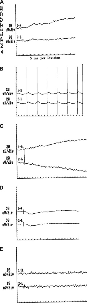
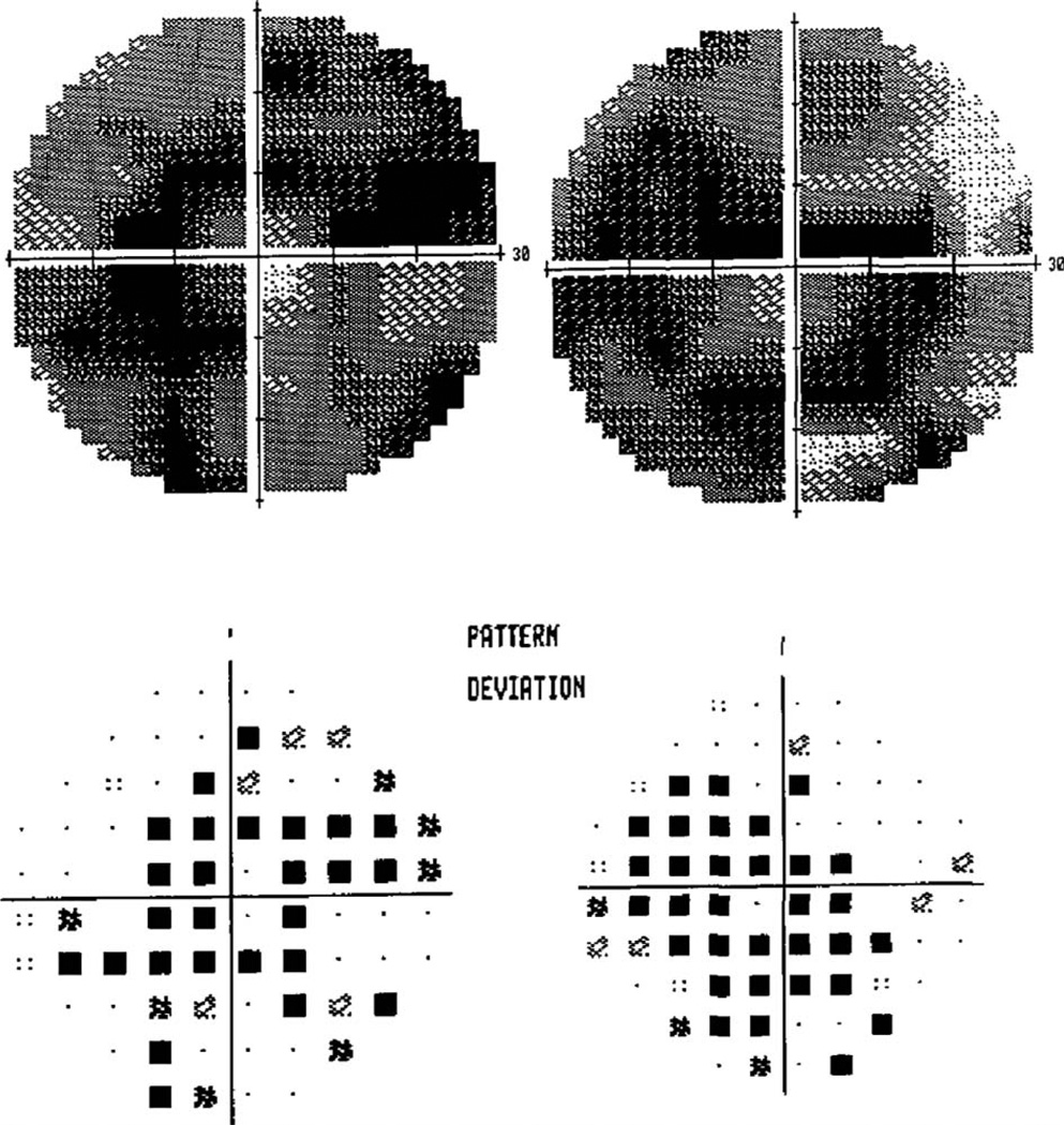
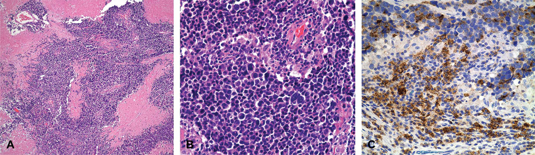
A 70-year-old woman developed progressive visual loss with compromised visual acuity and visual fields, cells in the anterior chamber and vitreous, attenuated retinal arterioles, and macular edema. She had undergone right oophorectomy and partial salpingectomy nearly 50 years earlier. Full-field and multifocal electroretinography showed waveforms of markedly attenuated amplitudes, findings consistent with cancer-associated retinopathy (CAR). Positron emission tomography revealed a nodule in the anterior wall of a right hydrosalpinx. Total laparoscopic hysterectomy yielded a neuroendocrine fallopian tube malignancy. She underwent partial treatment with paclitaxel and carboplatin that was aborted because of the development of herpes zoster infection. At 15 months following diagnosis, her ophthalmic status was stable. This is the first report of CAR in neuroendocrine carcinoma of the fallopian tube.
Figures
References
-
- Rosenblatt KA, Weiss NS, Schwartz SM. Incidence of malignant fallopian tube tumors. Incidence Gynecol Oncol. 1989;35:236–239. - PubMed
-
- Dursun P, Salman MC, Taskiran C, Usubutun A, Ayhan A. Primary neuroendocrine carcinoma of the fallopian tube: a case report. Am J Obstet Gynecol. 2004;190:568–571. - PubMed
-
- Bermudez A, Vighi S, Garcia A, Sardi J. Neuroendocrine cervical carcinoma: a diagnostic and therapeutic challenge. Gynecol Oncol. 2001;82:32–39. - PubMed
Publication types
MeSH terms
Substances
Grants and funding
LinkOut - more resources
Full Text Sources
Medical
