

Crystal structure of the alpha(6)beta(6) holoenzyme of propionyl-coenzyme A carboxylase
- PMID: 20725044
- PMCID: PMC2925307
- DOI: 10.1038/nature09302
Crystal structure of the alpha(6)beta(6) holoenzyme of propionyl-coenzyme A carboxylase
Abstract
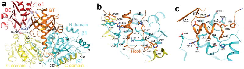
Propionyl-coenzyme A carboxylase (PCC), a mitochondrial biotin-dependent enzyme, is essential for the catabolism of the amino acids Thr, Val, Ile and Met, cholesterol and fatty acids with an odd number of carbon atoms. Deficiencies in PCC activity in humans are linked to the disease propionic acidaemia, an autosomal recessive disorder that can be fatal in infants. The holoenzyme of PCC is an alpha(6)beta(6) dodecamer, with a molecular mass of 750 kDa. The alpha-subunit contains the biotin carboxylase (BC) and biotin carboxyl carrier protein (BCCP) domains, whereas the beta-subunit supplies the carboxyltransferase (CT) activity. Here we report the crystal structure at 3.2-A resolution of a bacterial PCC alpha(6)beta(6) holoenzyme as well as cryo-electron microscopy (cryo-EM) reconstruction at 15-A resolution demonstrating a similar structure for human PCC. The structure defines the overall architecture of PCC and reveals unexpectedly that the alpha-subunits are arranged as monomers in the holoenzyme, decorating a central beta(6) hexamer. A hitherto unrecognized domain in the alpha-subunit, formed by residues between the BC and BCCP domains, is crucial for interactions with the beta-subunit. We have named it the BT domain. The structure reveals for the first time the relative positions of the BC and CT active sites in the holoenzyme. They are separated by approximately 55 A, indicating that the entire BCCP domain must translocate during catalysis. The BCCP domain is located in the active site of the beta-subunit in the current structure, providing insight for its involvement in the CT reaction. The structural information establishes a molecular basis for understanding the large collection of disease-causing mutations in PCC and is relevant for the holoenzymes of other biotin-dependent carboxylases, including 3-methylcrotonyl-CoA carboxylase (MCC) and eukaryotic acetyl-CoA carboxylase (ACC).
Figures


References
-
- Desviat LR, et al. Propionic acidemia: mutation update and functional and structural effects of the variant alleles. Mol Gen Metab. 2004;83:28–37. - PubMed
-
- Rodriguez-Pombo P, et al. Towards a model to explain the intragenic complementation in the heteromultimeric protein propionyl-CoA carboxylase. Biochim Biophys Acta. 2005;1740:489–498. - PubMed
-
- Deodato F, Boenzi S, Santorelli FM, Dionisi-Vici C. Methylmalonic and propionic aciduria. Am J Med Genet C Semin Med Genet. 2006;142:104–112. - PubMed
-
- Desviat LR, et al. New splicing mutations in propionic acidemia. J Hum Genet. 2006;51:992–997. - PubMed
-
- Desviat LR, et al. Functional analysis of MCCA and MCCB mutations causing methylcrotonylglycinuria. Mol Gen Metab. 2003;80:315–320. - PubMed
Publication types
MeSH terms
Substances
Associated data
- Actions
Grants and funding
LinkOut - more resources
Full Text Sources
Other Literature Sources
Molecular Biology Databases
Miscellaneous
