Review
. 2010 Sep;11(9):678-84.
doi: 10.1038/embor.2010.115. Epub 2010 Aug 20.

Mitochondrial shape changes: orchestrating cell pathophysiology

Affiliations
Review

Mitochondrial shape changes: orchestrating cell pathophysiology

Silvia Campello et al. EMBO Rep. 2010 Sep.

Abstract

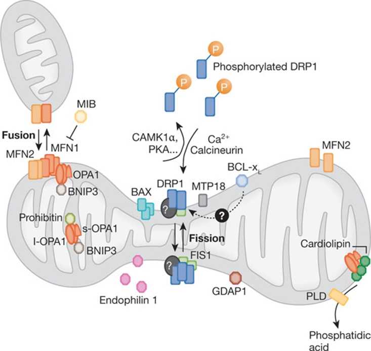
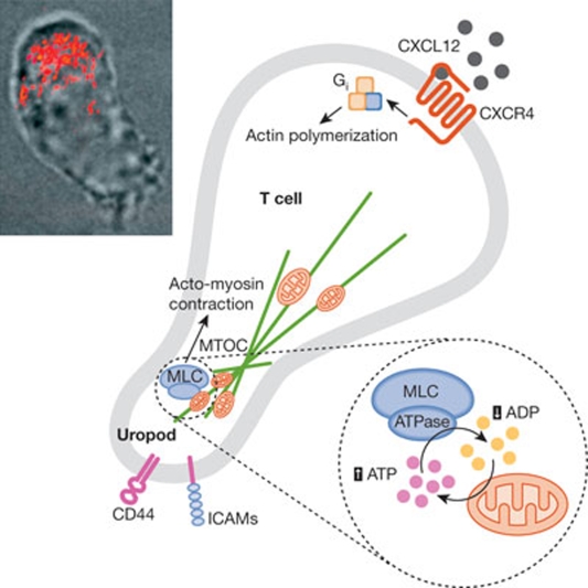
Mitochondria are highly dynamic organelles, the location, size and distribution of which are controlled by a family of proteins that modulate mitochondrial fusion and fission. Recent evidence indicates that mitochondrial morphology is crucial for cell physiology, as changes in mitochondrial shape have been linked to neurodegeneration, calcium signalling, lifespan and cell death. Because immune cells contain few mitochondria, these organelles have been considered to have only a marginal role in this physiological context-which is conversely well characterized from the point of view of signalling. Nevertheless, accumulating evidence shows that mitochondrial dynamics have an impact on the migration and activation of immune cells and on the innate immune response. Here, we discuss the roles of mitochondrial dynamics in cell pathophysiology and consider how studying dynamics in the context of the immune system could increase our knowledge about the role of dynamics in key signalling cascades.


Conflict of interest statement

The authors declare that they have no conflict of interest.

Figures

Figure 1
Figure 1
The dynamics of mitochondrial fission and fusion. The localization, as well as some interaction and modification of the principal proteins involved in the two processes are shown. Once dephosphorylated, DRP1 is recruited to the outer membrane by FIS1 or by another, unknown, component. The oligomerization of DRP1 is followed by constriction of the membrane and mitochondrial fission. The pro-fusion proteins (MFNs on the outer membrane and OPA1 on the inner membrane) oligomerize to induce fusion of the membranes. Other additional components of the machinery are shown. BAX, BCL2-associated X protein; BNIP3, BCL2/E1B 19 kDa-interacting protein 3; CAMK1a, calcium/calmodulin-dependent protein kinase 1a; DRP1, dynamin-related protein 1; FIS1, fission protein 1; GDAP1, ganglioside-induced differentiation-associated protein 1; l-OPA1, long form of OPA1; MFN, mitofusin; MIB, mitofusin-binding protein; MTP18, mitochondrial protein 18 kDa; OPA1, optic atrophy 1; PKA, protein kinase A; PLD, phospholipase D; s-OPA1: short form of OPA1.
Figure 2
Figure 2
A model for mitochondrial dynamics in the orchestration of T-cell chemotaxis. To achieve directed movement, lymphoid cells organize and maintain spatial and functional asymmetry, with a leading edge containing the machinery for actin polymerization and gradient sensing and a uropod containing the adhesion molecules and the MTOC. At the uropod, myosin filaments contract actin filaments to provide the tension required for cell movement. Myosin II activity is controlled mainly through the phosphorylation of MLC, which induces a conformational change allowing actin–myosin interactions and activating its ATPase activity. Myosin II ATPase depends on availability of the substrate and clearance of the product. Even in the presence of optimal ATP concentration, accumulation of ADP near the enzyme will slow down its activity and block migration. However, during lymphocyte migration, mitochondria are transported to the uropod along microtubules, in a process requiring Gi protein signalling and mitochondrial fission. The position of mitochondria is strategic for myosin ATPase activity: mitochondria not only supply ATP, but also withdraw ADP, thus creating optimal conditions for MLC. Inset: reconstruction of a confocal z-stack of images of a polarized Jurkat cell expressing mitochondrially targeted dsRED merged with a bright field image of the same cell showing polarization of mitochondria at the uropod. CXCL12, CXC ligand 12; CXCR4, CXC chemokine receptor 4; ICAM, intercellular adhesion molecule 1; MLC, myosin light chain; MTOC, microtubule organizing centre.
None
Silvia Campello
None
Luca Scorrano

References

    1. Abarca-Rojano E, Muniz-Hernandez S, Moreno-Altamirano MM, Mondragon-Flores R, Enriquez-Rincon F, Sanchez-Garcia FJ (2009) Re-organization of mitochondria at the NK cell immune synapse. Immunol Lett 122: 18–25 - PubMed
    1. Alexander C et al. (2000) OPA1, encoding a dynamin-related GTPase, is mutated in autosomal dominant optic atrophy linked to chromosome 3q28. Nat Genet 26: 211–215 - PubMed
    1. Autret A, Martin SJ (2009) Emerging role for members of the Bcl-2 family in mitochondrial morphogenesis. Mol Cell 36: 355–363 - PubMed
    1. Bellot G, Garcia-Medina R, Gounon P, Chiche J, Roux D, Pouyssegur J, Mazure NM (2009) Hypoxia-induced autophagy is mediated through hypoxia-inducible factor induction of BNIP3 and BNIP3L via their BH3 domains. Mol Cell Biol 29: 2570–2581 - PMC - PubMed
    1. Benard G, Rossignol R (2008) Mitochondrial fluidity matters. Focus on ‘Inherited complex I deficiency is associated with faster protein diffusion in the matrix of moving mitochondria'. Am J Physiol Cell Physiol 294: C1123 - PubMed

Publication types

MeSH terms

Substances