

The Nuclear Receptor PPARgamma as a Therapeutic Target for Cerebrovascular and Brain Dysfunction in Alzheimer's Disease
- PMID: 20725514
- PMCID: PMC2912024
- DOI: 10.3389/fnagi.2010.00021
The Nuclear Receptor PPARgamma as a Therapeutic Target for Cerebrovascular and Brain Dysfunction in Alzheimer's Disease
Abstract
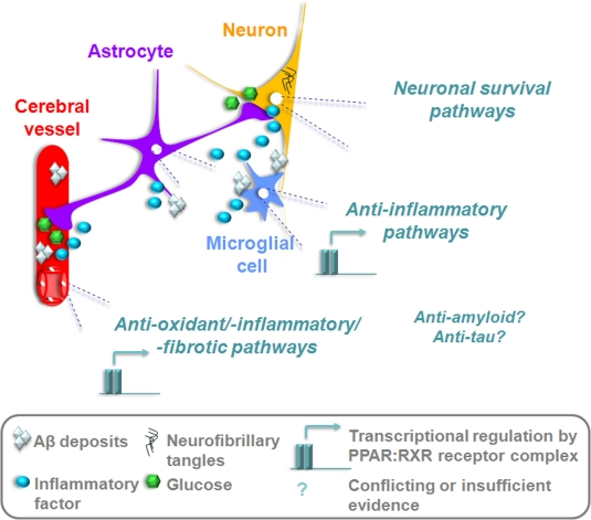
Peroxisome proliferator-activated receptors (PPARs) are ligand-activated nuclear transcription factors that regulate peripheral lipid and glucose metabolism. Three subtypes make up the PPAR family (alpha, gamma, beta/delta), and synthetic ligands for PPARalpha (fibrates) and PPARgamma (Thiazolidinediones, TZDs) are currently prescribed for the respective management of dyslipidemia and type 2 diabetes. In contrast to the well characterized action of PPARs in the periphery, little was known about the presence or function of these receptors in the brain and cerebral vasculature until fairly recently. Indeed, research in the last decade has uncovered these receptors in most brain cell types, and has shown that their activation, particularly that of PPARgamma, is implicated in normal brain and cerebrovascular physiology, and confers protection under pathological conditions. Notably, accumulating evidence has highlighted the therapeutic potential of PPARgamma ligands in the treatment of brain disorders such as Alzheimer's disease (AD), leading to the testing of the TZDs pioglitazone and rosiglitazone in AD clinical trials. This review will focus on the benefits of PPARgamma agonists for vascular, neuronal and glial networks, and assess the value of these compounds as future AD therapeutics in light of evidence from transgenic mouse models and recent clinical trials.
Keywords: arterial reactivity; brain metabolism; cerebral blood flow; inflammation; oxidative stress; pioglitazone; spatial memory; vascular fibrosis.
Figures
Similar articles
-
Peroxisome proliferator-activated receptors in vascular biology-molecular mechanisms and clinical implications.Vascul Pharmacol. 2006 Jul;45(1):19-28. doi: 10.1016/j.vph.2005.11.014. Epub 2006 Jun 16. Vascul Pharmacol. 2006. PMID: 16782410 Review.
-
Effects of peroxisome proliferator activated receptors (PPAR)-γ and -α agonists on cochlear protection from oxidative stress.PLoS One. 2017 Nov 28;12(11):e0188596. doi: 10.1371/journal.pone.0188596. eCollection 2017. PLoS One. 2017. PMID: 29182629 Free PMC article.
-
New approach in the treatment of T2DM and metabolic syndrome (focus on a novel insulin sensitizer).Acta Med Indones. 2006 Jul-Sep;38(3):160-6. Acta Med Indones. 2006. PMID: 17119268 Review.
-
Update on cardiovascular safety of PPARgamma agonists and relevance to medicinal chemistry and clinical pharmacology.Curr Top Med Chem. 2012;12(6):585-604. doi: 10.2174/156802612799436632. Curr Top Med Chem. 2012. PMID: 22242856 Review.
-
Peroxisome proliferator-activated receptors and atherogenesis: regulators of gene expression in vascular cells.Circ Res. 2004 May 14;94(9):1168-78. doi: 10.1161/01.RES.0000127122.22685.0A. Circ Res. 2004. PMID: 15142970 Review.
Cited by
-
Inhibition of PPARγ during rat pregnancy causes intrauterine growth restriction and attenuation of uterine vasodilation.Front Physiol. 2013 Jul 23;4:184. doi: 10.3389/fphys.2013.00184. eCollection 2013. Front Physiol. 2013. PMID: 23888144 Free PMC article.
-
Diabetes: Risk factor and translational therapeutic implications for Alzheimer's disease.Eur J Neurosci. 2022 Nov;56(9):5727-5757. doi: 10.1111/ejn.15619. Epub 2022 Feb 23. Eur J Neurosci. 2022. PMID: 35128745 Free PMC article.
-
Nuclear receptors in neurodegenerative diseases.Neurobiol Dis. 2014 Dec;72 Pt A:104-16. doi: 10.1016/j.nbd.2014.05.019. Epub 2014 May 27. Neurobiol Dis. 2014. PMID: 24874548 Free PMC article. Review.
-
Role of insulin resistance in Alzheimer's disease.Metab Brain Dis. 2015 Aug;30(4):839-51. doi: 10.1007/s11011-014-9631-3. Epub 2014 Nov 16. Metab Brain Dis. 2015. PMID: 25399337 Review.
-
Astragaloside IV, a Natural PPARγ Agonist, Reduces Aβ Production in Alzheimer's Disease Through Inhibition of BACE1.Mol Neurobiol. 2017 May;54(4):2939-2949. doi: 10.1007/s12035-016-9874-6. Epub 2016 Mar 29. Mol Neurobiol. 2017. PMID: 27023226
References
-
- Akiyama H., Barger S., Barnum S., Bradt B., Bauer J., Cole G. M., Cooper N. R., Eikelenboom P., Emmerling M., Fiebich B. L., Finch C. E., Frautschy S., Griffin W. S., Hampel H., Hull M., Landreth G., Lue L., Mrak R., Mackenzie I. R., McGeer P. L., O'Banion M. K., Pachter J., Pasinetti G., Plata-Salaman C., Rogers J., Rydel R., Shen Y., Streit W., Strohmeyer R., Tooyoma I., Van Muiswinkel F. L., Veerhuis R., Walker D., Webster S., Wegrzyniak B., Wenk G., Wyss-Coray T. (2000). Inflammation and Alzheimer's disease. Neurobiol. Aging 21, 383–42110.1016/S0197-4580(00)00124-X - DOI - PMC - PubMed
-
- Beyer A. M., Baumbach G. L., Halabi C. M., Modrick M. L., Lynch C. M., Gerhold T. D., Ghoneim S. M., de Lange W. J., Keen H. L., Tsai Y. S., Maeda N., Sigmund C. D., Faraci F. M. (2008). Interference with PPARγ signaling causes cerebral vascular dysfunction, hypertrophy and remodeling. Hypertension 51, 867–87110.1161/HYPERTENSIONAHA.107.103648 - DOI - PMC - PubMed
LinkOut - more resources
Full Text Sources