. 2010 Apr;3(4):24-38.

A review of acne in ethnic skin: pathogenesis, clinical manifestations, and management strategies

A review of acne in ethnic skin: pathogenesis, clinical manifestations, and management strategies

Erica C Davis et al. J Clin Aesthet Dermatol. 2010 Apr.

Abstract

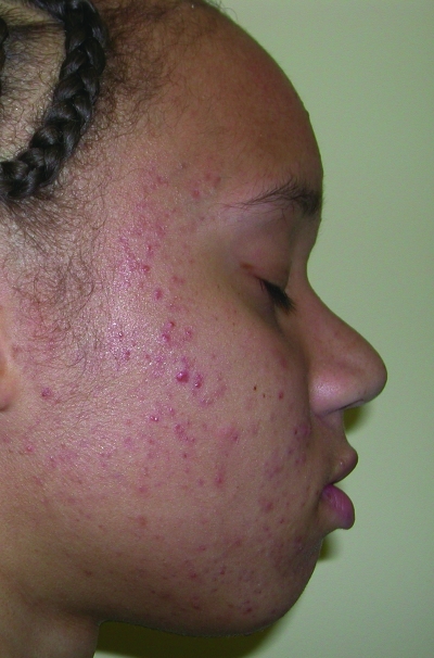
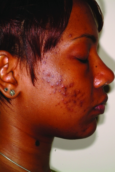
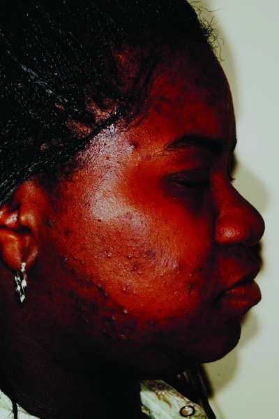
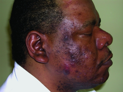
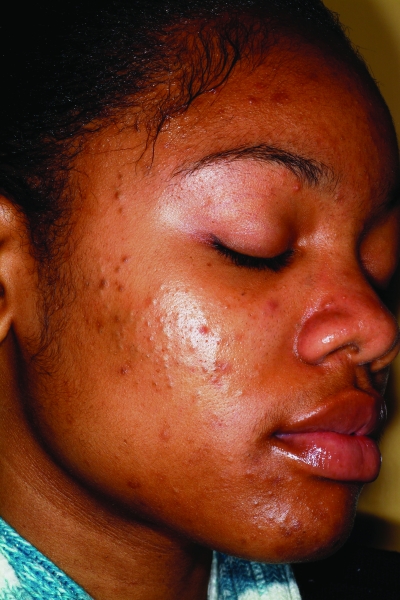
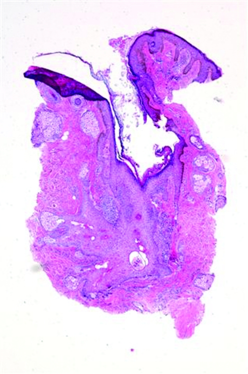
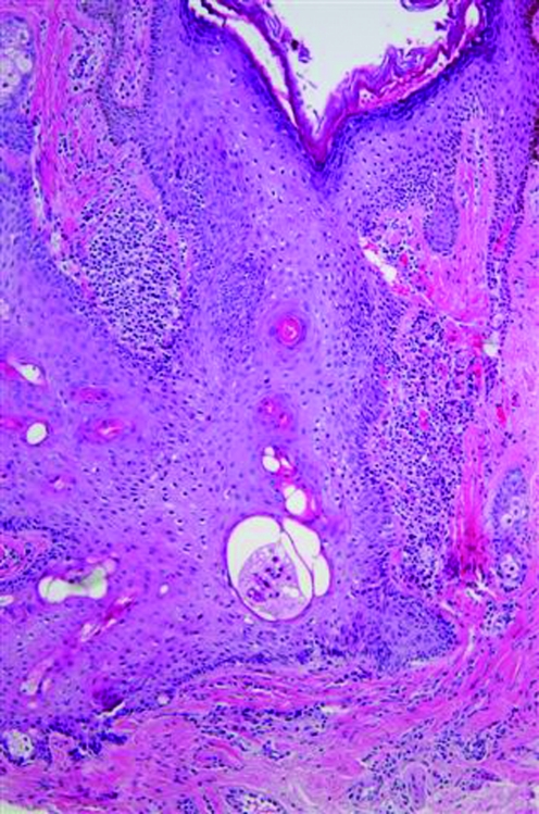
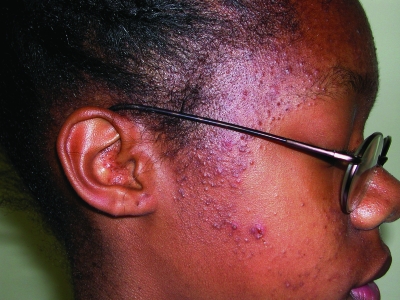
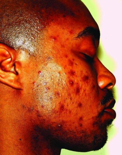
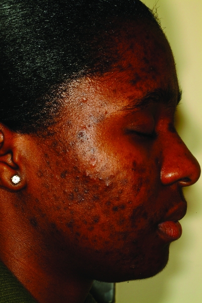
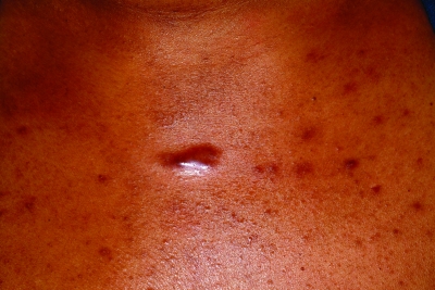
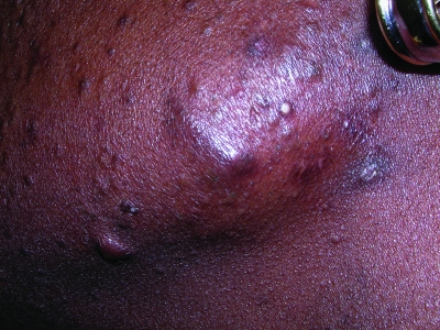
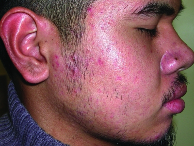
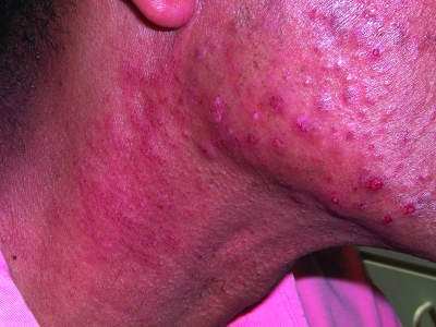
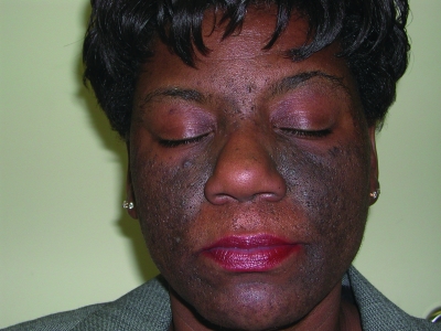
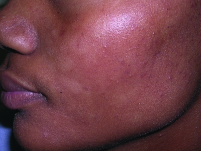
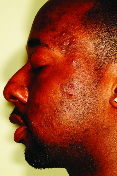
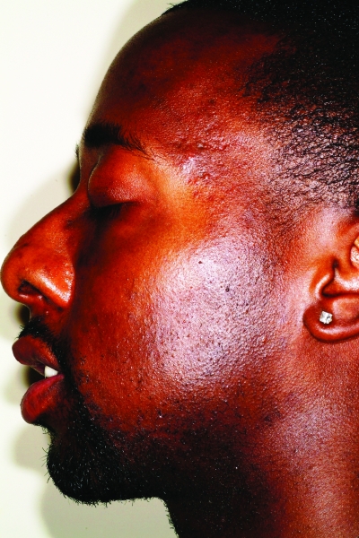
Acne vulgaris is one of the most common conditions for which all patients, including those with skin of color (Fitzpatrick skin types IV-VI), seek dermatological care. The multifactorial pathogenesis of acne appears to be the same in ethnic patients as in Caucasians. However, there is controversy over whether certain skin biology characteristics, such as sebum production, differ in ethnic patients. Clinically, acne lesions can appear the same as those seen in Caucasians; however, histologically, all types of acne lesions in African Americans can be associated with intense inflammation including comedones, which can also have some degree of inflammation. It is the sequelae of the disease that are the distinguishing characteristics of acne in skin of color, namely postinflammatory hyperpigmentation and keloidal or hypertrophic scarring. Although the medical and surgical treatment options are the same, it is these features that should be kept in mind when designing a treatment regimen for acne in skin of color.


Figures

Figures 1a–1c
Figures 1a–1c
Acne vulgaris in Fitzpatrick skin types IV (a), V (b), and VI (c). Note the postinflammatory hyperpigmentation, particularly in the higher skin phototypes.
Figures 1a–1c
Figures 1a–1c
Acne vulgaris in Fitzpatrick skin types IV (a), V (b), and VI (c). Note the postinflammatory hyperpigmentation, particularly in the higher skin phototypes.
Figures 1a–1c
Figures 1a–1c
Acne vulgaris in Fitzpatrick skin types IV (a), V (b), and VI (c). Note the postinflammatory hyperpigmentation, particularly in the higher skin phototypes.
Figure 2
Figure 2
Fitzpatrick skin type V patient with nodulocystic acne. Note the postinflammatory hyperpigmentation.
Figure 3
Figure 3
Comedonal acne. Note the presence of both open and closed comedones.
Figures 4a and 4b
Figures 4a and 4b
Histological examination of an open comedo. Note the dilated, distorted, keratin-filled follicle and the presence of patchy chronic inflammation. There was no evidence of inflammation on clinical exam.
Figures 4a and 4b
Figures 4a and 4b
Histological examination of an open comedo. Note the dilated, distorted, keratin-filled follicle and the presence of patchy chronic inflammation. There was no evidence of inflammation on clinical exam.
Figure 5
Figure 5
Patient with pomade acne eruption of closed comedones over the forehead and temple regions
Figures 6a and 6b
Figures 6a and 6b
Postinflammatory hyperpigmentation in a patient with Fitzpatrick skin type V (a) and VI (b). Note the greater intensity of pigmentation with darker skin
Figures 6a and 6b
Figures 6a and 6b
Postinflammatory hyperpigmentation in a patient with Fitzpatrick skin type V (a) and VI (b). Note the greater intensity of pigmentation with darker skin
Figures 7a and 7b
Figures 7a and 7b
Keloid formation on the chest (a) and along the mandible (b) from acne vulgaris
Figures 7a and 7b
Figures 7a and 7b
Keloid formation on the chest (a) and along the mandible (b) from acne vulgaris
Figure 8
Figure 8
Multiple areas of persistent erythema on the cheek after acne
Figures 9a, 9b, 9c
Figures 9a, 9b, 9c
Erythema (a), hyperpigmentation (b), hypopigmentation (c) resulting from an irritant contact dermatitis in skin of color patients. Figures 9b and 9c are reprinted with permission from Dermatologic Therapy. 2004;17(2):184–195.© John Wiley & Sons, Inc.
Figures 9a, 9b, 9c
Figures 9a, 9b, 9c
Erythema (a), hyperpigmentation (b), hypopigmentation (c) resulting from an irritant contact dermatitis in skin of color patients. Figures 9b and 9c are reprinted with permission from Dermatologic Therapy. 2004;17(2):184–195.© John Wiley & Sons, Inc.
Figures 9a, 9b, 9c
Figures 9a, 9b, 9c
Erythema (a), hyperpigmentation (b), hypopigmentation (c) resulting from an irritant contact dermatitis in skin of color patients. Figures 9b and 9c are reprinted with permission from Dermatologic Therapy. 2004;17(2):184–195.© John Wiley & Sons, Inc.
Figures 10a and 10b
Figures 10a and 10b
Skin of color patient with acne and PIH before (a) and after (b) 7 months of therapy with tazarotene 0.1% applied in the mornings and a fixed combination lightening agent (fluocinolone acetonide 0.01%, hydroquinone 4%, and tretinoin 0.05% cream) applied in the evenings
Figures 10a and 10b
Figures 10a and 10b
Skin of color patient with acne and PIH before (a) and after (b) 7 months of therapy with tazarotene 0.1% applied in the mornings and a fixed combination lightening agent (fluocinolone acetonide 0.01%, hydroquinone 4%, and tretinoin 0.05% cream) applied in the evenings

Similar articles

Cited by

References

    1. White GM. Recent findings in the epidemiologic evidence, classification, and subtypes of acne vulgaris. J Am Acad Dermatol. 1998;39:S34–S37. - PubMed
    1. Alexis AF, Sergay AB, Taylor SC. Common dermatologic disorders in skin of color: a comparative practice survey. Cutis. 2007;80:38–394. - PubMed
    1. Fox H. Observations on skin diseases in the Negro. J Cutan Dis. 1908;26:67–79.
    1. Hazen HH. Personal observations upon skin diseases in the American Negro. J Cutan Dis. 1914;32:705–712.
    1. Hazen HH. Syphilis and skin disease in the American Negro: personal observations. AMA Arch Derm Syphilol. 1935;31:316–323.

LinkOut - more resources