Spike timing dependent plasticity: a consequence of more fundamental learning rules
- PMID: 20725599
- PMCID: PMC2922937
- DOI: 10.3389/fncom.2010.00019
Spike timing dependent plasticity: a consequence of more fundamental learning rules
Abstract
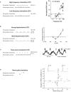
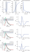
Spike timing dependent plasticity (STDP) is a phenomenon in which the precise timing of spikes affects the sign and magnitude of changes in synaptic strength. STDP is often interpreted as the comprehensive learning rule for a synapse - the "first law" of synaptic plasticity. This interpretation is made explicit in theoretical models in which the total plasticity produced by complex spike patterns results from a superposition of the effects of all spike pairs. Although such models are appealing for their simplicity, they can fail dramatically. For example, the measured single-spike learning rule between hippocampal CA3 and CA1 pyramidal neurons does not predict the existence of long-term potentiation one of the best-known forms of synaptic plasticity. Layers of complexity have been added to the basic STDP model to repair predictive failures, but they have been outstripped by experimental data. We propose an alternate first law: neural activity triggers changes in key biochemical intermediates, which act as a more direct trigger of plasticity mechanisms. One particularly successful model uses intracellular calcium as the intermediate and can account for many observed properties of bidirectional plasticity. In this formulation, STDP is not itself the basis for explaining other forms of plasticity, but is instead a consequence of changes in the biochemical intermediate, calcium. Eventually a mechanism-based framework for learning rules should include other messengers, discrete change at individual synapses, spread of plasticity among neighboring synapses, and priming of hidden processes that change a synapse's susceptibility to future change. Mechanism-based models provide a rich framework for the computational representation of synaptic plasticity.
Keywords: STDP; calcium; learning rules; long-term depression; long-term potentiation; mechanistic models; synaptic plasticity.
Figures


Similar articles
-
Interaction of inhibition and triplets of excitatory spikes modulates the NMDA-R-mediated synaptic plasticity in a computational model of spike timing-dependent plasticity.Hippocampus. 2013 Jan;23(1):75-86. doi: 10.1002/hipo.22057. Epub 2012 Jul 31. Hippocampus. 2013. PMID: 22851353
-
Temporal modulation of spike-timing-dependent plasticity.Front Synaptic Neurosci. 2010 Jun 17;2:19. doi: 10.3389/fnsyn.2010.00019. eCollection 2010. Front Synaptic Neurosci. 2010. PMID: 21423505 Free PMC article.
-
Synaptic plasticity rules with physiological calcium levels.Proc Natl Acad Sci U S A. 2020 Dec 29;117(52):33639-33648. doi: 10.1073/pnas.2013663117. Epub 2020 Dec 16. Proc Natl Acad Sci U S A. 2020. PMID: 33328274 Free PMC article.
-
Dendritic mechanisms controlling spike-timing-dependent synaptic plasticity.Trends Neurosci. 2007 Sep;30(9):456-63. doi: 10.1016/j.tins.2007.06.010. Epub 2007 Aug 31. Trends Neurosci. 2007. PMID: 17765330 Review.
-
A Hypothetical Model Concerning How Spike-Timing-Dependent Plasticity Contributes to Neural Circuit Formation and Initiation of the Critical Period in Barrel Cortex.J Neurosci. 2019 May 15;39(20):3784-3791. doi: 10.1523/JNEUROSCI.1684-18.2019. Epub 2019 Mar 15. J Neurosci. 2019. PMID: 30877173 Free PMC article. Review.
Cited by
-
Role of sensory input distribution and intrinsic connectivity in lateral amygdala during auditory fear conditioning: a computational study.Neuroscience. 2012 Nov 8;224:249-67. doi: 10.1016/j.neuroscience.2012.08.030. Epub 2012 Aug 21. Neuroscience. 2012. PMID: 22917618 Free PMC article.
-
Functional mechanisms underlie the emergence of a diverse range of plasticity phenomena.PLoS Comput Biol. 2018 Nov 12;14(11):e1006590. doi: 10.1371/journal.pcbi.1006590. eCollection 2018 Nov. PLoS Comput Biol. 2018. PMID: 30419014 Free PMC article.
-
Theoretical analysis of low power optogenetic control of synaptic plasticity with subcellular expression of CapChR2 at postsynaptic spine.Sci Rep. 2025 Apr 1;15(1):11166. doi: 10.1038/s41598-025-95355-6. Sci Rep. 2025. PMID: 40169824 Free PMC article.
-
Dynamic stability of sequential stimulus representations in adapting neuronal networks.Front Comput Neurosci. 2014 Oct 22;8:124. doi: 10.3389/fncom.2014.00124. eCollection 2014. Front Comput Neurosci. 2014. PMID: 25374534 Free PMC article.
-
Heterogeneous quantization regularizes spiking neural network activity.Sci Rep. 2025 Apr 23;15(1):14045. doi: 10.1038/s41598-025-96223-z. Sci Rep. 2025. PMID: 40268966 Free PMC article.
References
Grants and funding
LinkOut - more resources
Full Text Sources
Other Literature Sources
Miscellaneous

