. 2010 Jul 27:2010:260512.
doi: 10.4061/2010/260512.

DNA mismatch repair in eukaryotes and bacteria

Affiliations

DNA mismatch repair in eukaryotes and bacteria

Kenji Fukui. J Nucleic Acids. .

Abstract

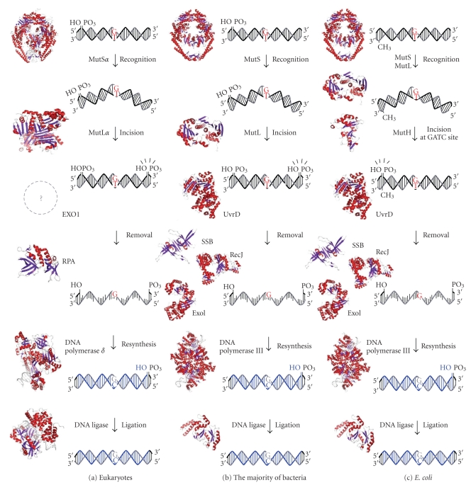
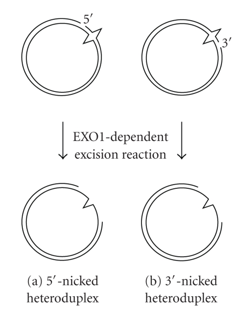
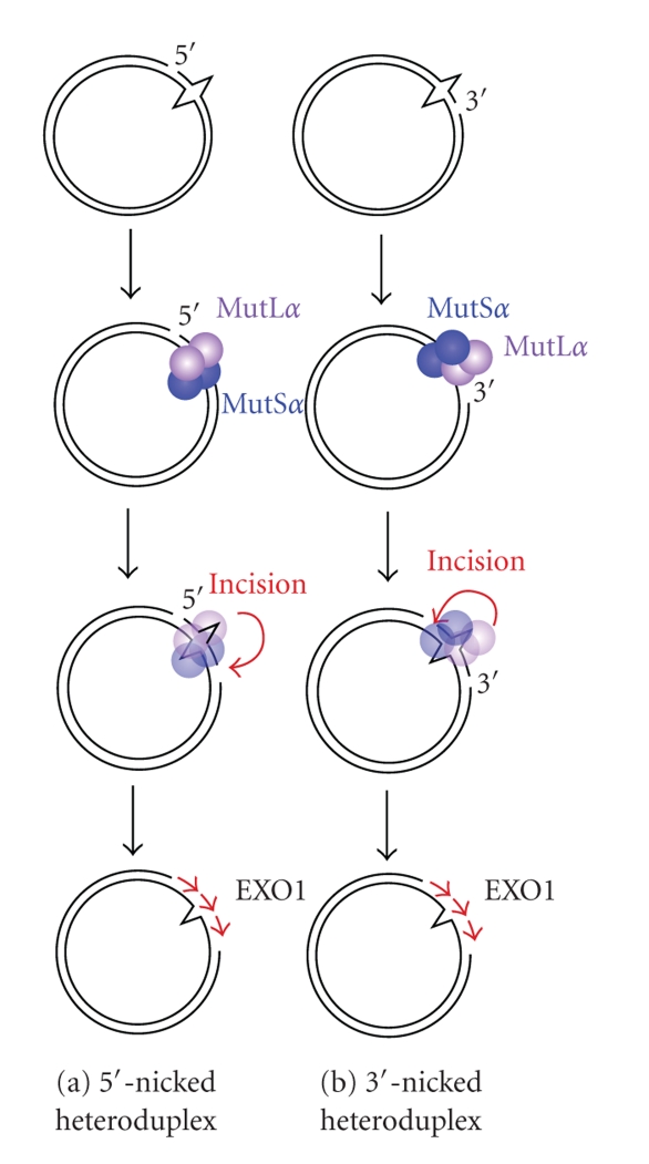
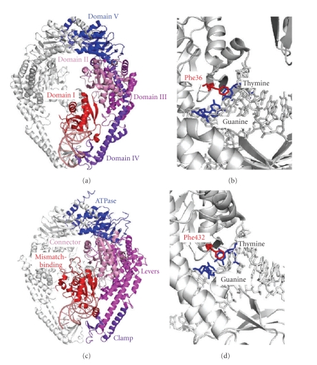
DNA mismatch repair (MMR) corrects mismatched base pairs mainly caused by DNA replication errors. The fundamental mechanisms and proteins involved in the early reactions of MMR are highly conserved in almost all organisms ranging from bacteria to human. The significance of this repair system is also indicated by the fact that defects in MMR cause human hereditary nonpolyposis colon cancers as well as sporadic tumors. To date, 2 types of MMRs are known: the human type and Escherichia coli type. The basic features of the former system are expected to be universal among the vast majority of organisms including most bacteria. Here, I review the molecular mechanisms of eukaryotic and bacterial MMR, emphasizing on the similarities between them.


Figures

Figure 1
Figure 1
A schematic representation of MMR pathway models. (a) Eukaryotic MMR. A DNA mismatch is generated by the misincorporation of a base during DNA replication. MutSα recognizes base-base mismatches and MutLα nicks the 3′- or 5′-side of the mismatched base on the discontinuous strand. The resulting DNA segment is excised by the EXO1 exonuclease, in cooperation with the single-stranded DNA-binding protein RPA. The DNA strand is resynthesized by DNA polymerase δ and DNA ligase 1. (b) MMR in mutH-less bacteria. Mismatched bases are recognized by MutS. After the incision of discontinuous strand by MutL, the error-containing DNA strand is removed by the cooperative functions of DNA helicases, such as UvrD, the exonucleases RecJ and ExoI, and the single-stranded DNA-binding protein SSB. DNA polymerase III and DNA ligase fill the gap to complete the repair. (c) E. coli MMR. MutS recognizes mismatched bases, and MutL interacts with and stabilizes the complex. Then, MutH endonuclease is activated to incise the unmethylated GATC site to create an entry point for the excision reaction. DNA helicase, a single-stranded DNA-binding protein, and several exonucleases are involved in the excision reaction. PDB IDs of crystal structures in this figure are 2O8B (human MutSα), 1H7S (human MutLα), 1L1O (human RPA), 3IAY (human DNA polymerase δ), 1X9N (human DNA ligase 1), 1E3M (bacterial MutS), 1B63 (bacterial MutL), 2AZO (E. coli MutH), 2ISI (bacterial UvrD), 2ZXO (bacterial RecJ), 3C95 (bacterial ExoI), 2CWA (bacterial SSB), 2HQA (bacterial DNA polymerase III), and 2OWO (bacterial DNA ligase).
Figure 2
Figure 2
Bidirectionality of eukaryotic MMR. The 5′-nicked (a) and 3′-nicked (b) heteroduplexes are used as model substrate. The shorter path is chosen to remove the mismatched base. The 5′–3′ exonuclease activity of EXO1 is required for excision reaction in both 5′- and 3′-nicked heteroduplexes.
Figure 3
Figure 3
The 5′-nick-(a) and 3′-nick-(b) directed eukaryotic MMR. After recognition of a mismatched base by MutSα, MutLα incises the discontinuous strand of the heteroduplex in a mismatch-MutSα-, PCNA-, RFC-, and ATP-dependent manner [10, 30, 31]. Incisions by MutLα occur dominantly on the distal side of the mismatched base relative to the pre-existed strand break although it can also occur proximal to the mismatch [30].
Figure 4
Figure 4
Crystal structures of MutS-mismatch complex. (a) Crystal structure of E. coli MutS bound to a G:T-mismatched heteroduplex (PDB ID: 1E3M). One of the 2 subunits of E. coli MutS is shown in color. DNA is shown in salmon. Domains I, II, III, IV, and V are shown in red, pink, violet, purple blue, and blue, respectively. Domains I, IV, and V are responsible for mismatch recognition, double-stranded DNA binding, and dimerization/ATP binding, respectively. (b) The mismatch-recognition site in E. coli MutS-G:T mismatch complex. The mismatch-recognizing phenylalanine residue (Phe36) and G:T mismatch are shown in red and blue, respectively. (c) Crystal structure of human MutSα (PDB ID: 2O8B), which is comprised of full-length MSH2 and a protease-resistant fragment of MSH6 lacking the first 340 amino acid residues. MSH2 is shown in white and MSH6 is in color. Mismatch-binding, Connector, Levers, Clamp, and ATPase domains are colored in red, pink, magenta, purple, and blue, respectively. (d) The mismatch-recognition site in human MutSα-G:T mismatch complex. Phe432 and G:T mismatch is shown in red and blue, respectively.
Figure 5
Figure 5
Mismatch recognition mode of MutS. (a) G:T mismatch bound to E. coli MutS (PDB ID: 1E3M). (b) C:A mismatch bound to E. coli MutS (PDB ID: 1OH5). Cytosine residue is in a syn conformation. (c) G:T mismatch bound to human MutSα (PDB ID: 2O8B). (d) Side view of the E. coli MutS-mismatch complex (PDB ID: 1E3M). The mismatched duplex is sharply kinked in the complex with MutS. MutS and mismatched DNA are colored grey and red, respectively. The mismatched G and T are shown in the sphere model.
Figure 6
Figure 6
The model for full length of E. coli MutS dimer. The crystal structure of the C-terminal 34 amino acids of E. coli MutS (2OK2) [44] was connected to the C-termini of E. coli MutS (residues 2-800) structure (1E3M).
Figure 7
Figure 7
(a) A schematic representation of the domain structure of MutL homologues. ATPase, nuclease, and dimer indicate the ATPase, endonuclease, and dimerization domains, respectively. The crystal structures of N-terminal ATPase domain of human PMS2 (PDB ID: 1EA6) [45], ATPase domain of E. coli MutL (PDB ID: 1B63) [46], and C-terminal dimerization domain of E. coli MutL (PDB ID: 1X9Z) [47, 48] are shown.
Figure 8
Figure 8
Crystal structures of the apo form ((a) PDB ID: 1BKN) and ADPNP-bound form ((b) PDB ID: 1B63) of the E. coli MutL N-terminal domain. (c) A schematic representation of a model for the ATP-dependent conformational change of full-length MutLα [73]. NTD and CTD indicate the N-terminal ATPase domain and the C-terminal endonuclease domain, respectively. In the apo form of MutLα, the PMS2 and MLH1 subunits dimerize via their C-terminal domains. ATP binding induces the dimerization of the N-terminal domain and condensation of the molecule.
Figure 9
Figure 9
A model for the ATPase-cycle-dependent regulation of bacterial MutL endonuclease activity. Free MutL exists as an ATP-bound form whose endonuclease activity is inactive, but preferably binds to a MutS-mismatch complex. The interaction with the MutS-mismatch complex induces the ATP hydrolysis of MutL, resulting in the stimulation of its endonuclease activity. Adapted from the work of Fukui et al. [34].
Figure 10
Figure 10
Two parallel pathways of excision reaction in T. thermophilus. RecJ (red) and ExoI (purple) are thought to be responsible for the 5′- and 3′-directed excision, respectively. UvrD helicase (magenta) functions in cooperation with RecJ. DNA helicase (blue) which translocates 5′ to 3′ direction has been unknown.
Figure 11
Figure 11
Crystal structure of T. thermophilus RecJ (PDB ID: 2ZXR). Full-length T. thermophilus RecJ is comprised of domains I–IV and forms a ring-like structure. The catalytic active site is located in the cavity between domains I and II as indicated by an arrow. Domain III shows a structural similarity to the oligonucleotide/oligosaccharide-binding fold that is often found in single-stranded DNA-binding proteins. The ring-like structure and oligonucleotide/oligosaccharide-binding fold will ensure the high processivity and strict specificity for single-stranded DNA.
Figure 12
Figure 12
Prevention of 8OG-induced G:C-T:A transversion mutations. The 8OG base is one of the major forms of oxidative DNA damage and can be generated by reactive oxygen species. Since 8OG can pair not only with C but also with A, it causes a G:C-T:A transversion through DNA replication. MutM (OGG1)- and MutY (MYH)-dependent base-excision repair pathways are known to remove the 8OG and A from 8OG:C and 8OG:A pairs, respectively. MMR is also responsible for the removal of A from 8OG.

References

    1. Friedberg EC, Walker GC, Siede W. DNA Repair and Mutagenesis. 2nd edition. Washington, DC, USA: American Society for Microbiology; 2005.
    1. Schaaper RM. Base selection, proofreading, and mismatch repair during DNA replication in Escherichia coli . Journal of Biological Chemistry. 1993;268(32):23762–23765. - PubMed
    1. Fishel R, Kolodner RD. Identification of mismatch repair genes and their role in the development of cancer. Current Opinion in Genetics and Development. 1995;5(3):382–395. - PubMed
    1. Suter CM, Martin DIK, Ward RL. Germline epimutation of MLH1 in individuals with multiple cancers. Nature Genetics. 2004;36(5):497–501. - PubMed
    1. Kane MF, Loda M, Gaida GM, et al. Methylation of the hMLH1 promoter correlates with lack of expression of hMLH1 in sporadic colon tumors and mismatch repair-defective human tumor cell lines. Cancer Research. 1997;57(5):808–811. - PubMed

LinkOut - more resources