Review
. 2010 Nov;101(11):2302-8.
doi: 10.1111/j.1349-7006.2010.01687.x.

Neogenesis and development of the high endothelial venules that mediate lymphocyte trafficking

Affiliations
Review

Neogenesis and development of the high endothelial venules that mediate lymphocyte trafficking

Haruko Hayasaka et al. Cancer Sci. 2010 Nov.

Abstract

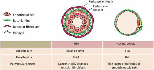
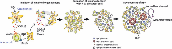
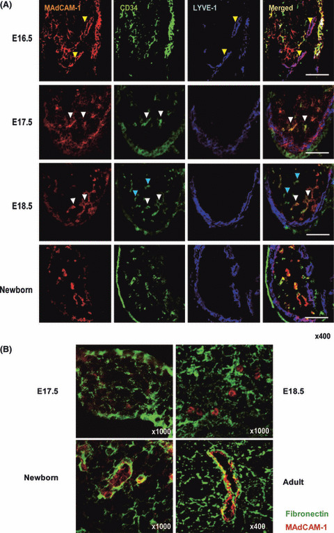
Physiological recruitment of lymphocytes from the blood into lymph nodes and Peyer's patches is mediated by high endothelial venules (HEV), specialized blood vessels found in secondary lymphoid tissues except for the spleen. The HEV are distinguished from other types of blood vessels by their tall and plump endothelial cells, and by their expression of specific chemokines and adhesion molecules, which all contribute to the selective lymphocyte trafficking across these blood vessels. The development of HEV is ontogenically regulated, and they appear perinatally in the mouse. High endothelial venules can appear ectopically, for instance in chronically inflamed tissues. Given that HEV enable the efficient trafficking of lymphocytes into tissues, the induction of HEV at a tumor site could potentiate tumor-specific immune responses, and the artificial manipulation of HEV neogenesis might thus provide a new tool for cancer immunotherapy. However, the process of HEV development and the mechanisms by which the unique features of HEV are maintained are incompletely understood. In this review, we discuss the process of HEV neogenesis and development during ontogeny, and their molecular requirements for maintaining their unique characteristics under physiological conditions.


Figures

Figure 1
Figure 1
The unique morphological features of high endothelial venules (HEV). The HEV are easily distinguished from normal venules by their specialized endothelial cells, the high endothelial cells, which have a tall and plump shape. A single layer of high endothelial cells is surrounded by thick basal lamina composed of fibronectin, collagen IV, and laminins. The HEV are further enclosed by concentric layers of fibroblastic reticular cells.
Figure 2
Figure 2
Schematic of lymphoid tissue organogenesis and high endothelial venule (HEV) development. Lymphoid organogenesis starts with expression of CXCL13 from stromal inducer cells in response to retinoic acid (RA) produced by adjacent cells, such as neurons. CXCL13 attract CXCR5+ LTα1β2 + lymphoid tissue inducer cells, which leads to the triggering of LTβR on stromal organizer cells. The stimulated organizer cells produce the chemokines CXCL13, CCL19 and CCL21, resulting in further accumulation of lymphoid inducer cells. After stable cell clustering occurs, HEV precursors differentiate into mature HEV, induced by interactions with the lymphoid tissue microenvironment.
Figure 3
Figure 3
Histological demonstration of high endo‐thelial venule (HEV) neogenesis. (A) In 16.5‐dpc mesenteric lymph nodes (MLN), MAdCAM‐1+ vascular structures that express LYVE‐1 but not CD34 are observed (yellow arrowheads), indicating that lymphatic vessel formation starts at this stage. From 17.5 dpc onward, most MAdCAM‐1+ structures are CD34+ (white arrowheads). No blood vessels with a distinct lumen are observed at this time. In 18.5‐dpc MLNs, MAdCAM‐1+/CD34+ (white arrowheads) and MAdCAM‐1/CD34+ (blue arrowheads) cell aggregates are observed; HEV‐type and non‐HEV‐type blood vessels appear at this stage. At this stage, LYVE‐1 expression is more prominent in the subcapsular region, whereas MAdCAM‐1 expression is less prominent. In newborn MLN, the expression of MAdCAM‐1 and LYVE‐1 are geographically segregated. Bars, 100 μm. (B) At 17.5 and 18.5 dpc, the expression of fibronectin (green) is scattered, and not necessarily closely associated with MAdCAM‐1+ cell aggregates (red). In newborn MLN, the basal lamina is prominent around MAdCAM‐1+ structures, sugges‐ting that the fibronectin‐expressing basal lamina typically seen in HEV develops prenatally.

Similar articles

Cited by

References

    1. Von Andrian UH, Mempel TR. Homing and cellular traffic in lymph nodes. Nat Rev Immunol 2003; 3: 867–78. - PubMed
    1. Miyasaka M, Tanaka T. Lymphocyte trafficking across high endothelial venules: dogmas and enigmas. Nat Rev Immunol 2004; 4: 360–70. - PubMed
    1. Bajenoff M, Egen JG, Koo LY et al. Stromal cell networks regulate lymphocyte entry, migration, and territoriality in lymph nodes. Immunity 2006; 25: 989–1001. - PMC - PubMed
    1. Mebius RE, Dowbenko D, Williams A, Fennie C, Lasky LA, Watson SR. Expression of GlyCAM‐1, an endothelial ligand for L‐selectin, is affected by afferent lymphatic flow. J Immunol 1993; 151: 6769–76. - PubMed
    1. Mebius RE, Breve J, Duijvestijn AM, Kraal G. The function of high endothelial venules in mouse lymph nodes stimulated by oxazolone. Immunology 1990; 71: 423–7. - PMC - PubMed