. 2010 Dec;161(8):1708-21.
doi: 10.1111/j.1476-5381.2010.00994.x.

Pioglitazone attenuates prostatic enlargement in diet-induced insulin-resistant rats by altering lipid distribution and hyperinsulinaemia

Affiliations

Pioglitazone attenuates prostatic enlargement in diet-induced insulin-resistant rats by altering lipid distribution and hyperinsulinaemia

Ajit Vikram et al. Br J Pharmacol. 2010 Dec.

Abstract

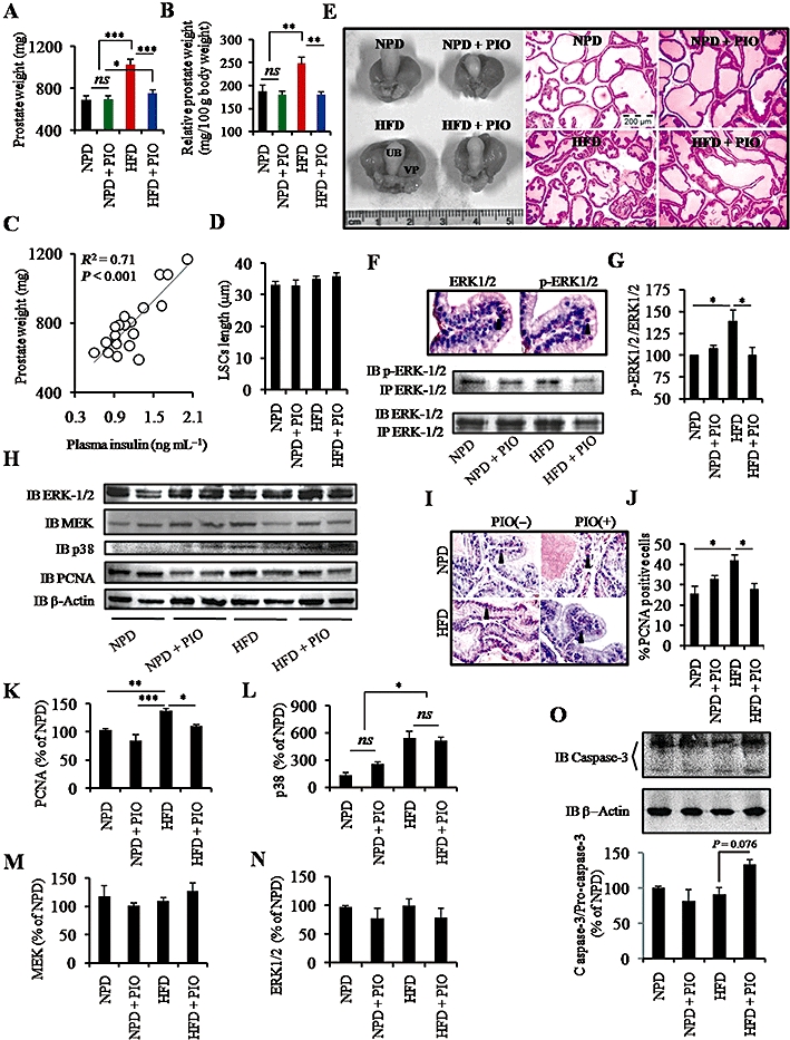
Background and purpose: Increased incidence of benign prostatic hyperplasia among insulin-resistant individuals suggests a role for hyperinsulinaemia in prostatic enlargement. We have already reported increased cell proliferation and enlargement of prostate gland in insulin-resistant rats. The present study aimed to elucidate the molecular mechanisms underlying the reversal of prostatic enlargement in insulin-resistant rats by the peroxisome proliferator-activated receptor γ agonist pioglitazone.

Experimental approach: Sprague-Dawley rats were fed a normal pellet or a high-fat diet for 12 weeks with or without pioglitazone (20 mg·kg(-1)). Subgroups of animals fed different diets were castrated. Effects of dietary manipulation and pioglitazone were measured on insulin sensitivity, lipid distribution, cell proliferation and apoptosis.

Key results: A high-fat diet led to the accumulation of fat in non-adipose tissues, insulin resistance, compensatory hyperinsulinaemia and prostatic enlargement in rats. Pioglitazone treatment altered fat distribution, improved insulin sensitivity and normalized lipid and insulin level in rats on the high-fat diet. The improved metabolic parameters led to decreased cellular proliferation and increased apoptosis in the prostate gland. High-fat diet feeding and pioglitazone treatment did not change plasma testosterone levels. However, significant prostatic atrophy was observed in castrated rats irrespective of dietary intervention.

Conclusions and implications: Our results show a previously unexplored therapeutic potential of pioglitazone for prostatic enlargement under insulin-resistant condition and further suggest that targeting distribution of lipid from non-adipose tissue to adipose tissue and insulin signalling could be new strategies for the treatment of benign prostatic hyperplasia.


Figures

Figure 1
Figure 1
Pioglitazone treatment (PIO) restores normoglycaemia and normoinsulinaemia in insulin-resistant rats and improves insulin sensitivity. (A) Body weight was increased in high-fat diet (HFD)-fed rats, compared to the age-matched control (n = 5–6). (B,C) Pioglitazone treatment l decreased the plasma glucose (B) and insulin (C) level in the HFD-fed rats, compared to diet-matched controls (n = 5). (D) No change in the plasma testosterone level was observed among the different groups (n = 4). (E) The adiposity index was increased in HFD-fed rats and pioglitazone treatment further increased the adiposity (n = 5). (F–I) Pioglitazone partially improved HFD induced glucose intolerance (glucose tolerance test, GTT) and impairment in the insulin mediated glucose disposal (insulin tolerance test, ITT) as shown by changes in the area under curve (AUC) (n = 4). All the values are shown as mean ± SEM. *P < 0.05, **P < 0.01, ***P < 0.001 and ns (not significant) P > 0.05. NPD, normal pellet diet.
Figure 2
Figure 2
Pioglitazone redirects lipid to adipose tissue and improves lipid levels. (A,B) Pioglitazone treatment (PIO) decreased the plasma cholesterol (A) and triglyceride (B) levels in the high-fat diet (HFD)-fed rats compared to the diet-matched control. Although there were no significant differences between HDL-cholesterol level among all the groups, the HDL-cholesterol level tended to increase in the HFD + PIO group, compared to HFD-fed rats (n = 5). LDL-cholesterol was decreased in HFD + PIO group as compared to the HFD-fed rats. (C) HFD feeding significantly increased liver weight (n = 5). (D,E) Pioglitazone treatment decreased in the liver triglyceride (D) and cholesterol (E) level in the HFD-fed rats compared to the diet-matched control (n = 5). (F) Pioglitazone treatment marginally decreased the muscle triglyceride level in the HFD-fed rats compared to diet-matched controls (n = 3–5). (G) Representative photomicrographs showing effects of dietary manipulation and pioglitazone treatment on liver, muscle, brown adipose tissue (BAT) and white adipose tissue (WAT) at different magnifications. Panels a–c show excessive fat deposition in the hepatocytes and subsequent normalization with pioglitazone treatment. Panel d shows the histological sections of skeletal muscle (SM). Panels e–g show the enlargement of adipocytes of WAT in HFD-fed rats and normalization with pioglitazone treatment. Panels h–j shows the enlargement of adipocytes of WAT in HFD-fed rats and normalization with pioglitazone treatment. In BAT of HFD-fed rats receiving pioglitazone treatment, several foci of small adipocytes were observed and the representative photomicrographs were shown at different magnification. All the values are shown as mean ± SEM. *P < 0.05, **P < 0.01, ***P < 0.001 and ns (not significant) P > 0.05 versus indicated group, ††P < 0.01, †††P < 0.001 versus indicated group for LDL-cholesterol. HDL, high-density lipoprotein; LDL, low-density lipoprotein; NPD, normal pellet diet.
Figure 3
Figure 3
Pioglitazone restores Hyperinsulinaemia-induced augmented cell proliferation and prostatic enlargement. (A,B) High-fat diet (HFD) feeding increased in the absolute (A) and relative (B) prostate weight and pioglitazone treatment (PIO) inhibited the diet-induced prostatic enlargement (n = 10). (C) Correlation between plasma insulin level and prostate weight. (D) HFD feeding and subsequent pioglitazone treatment did not affect the cellular height and morphology of the luminal secretory epithelial cells of the ventral prostate (n = 4). (E) Representative photomicrographs showing prostatic enlargement in the HFD-fed rats and restoration with the pioglitazone treatment. Increased infolding of the epithelial layer was observed in HFD-fed rats, compared to normal pellet diet.(NPD)-fed rats. (F,G) Effects of dietary manipulation and pioglitazone treatment on the activation of extracellular signal-regulated kinase (ERK1/2). Photomicrographs showing immunohistochemical localization of ERK1/2 and phosphorylated ERK 1/2 (p-ERK1/2) in the nucleus of prostatic luminal secretory epithelial cells and Western blot analysis of immunoprecipitated ERK for phosphorylation (E). Significantly increased p-ERK1/2 : ERK1/2 ratio was observed in the HFD-fed hyperinsulinaemic rats. Pioglitazone treatment restored the p-ERK1/2 : ERK1/2 ratio in HFD-fed rats (F) (n = 3). (H) Immunoblots of different proteins [β-Actin, proliferating cell nuclear antigen (PCNA), p38, mitrogen-activated protein kinase kinase (MEK) and ERK1/2]. (I) Immunostaining of the prostatic sections of different groups for PCNA. (J) A decrease in cells immunopositive for PCNA was observed in the HFD-fed rats receiving pioglitazone treatment compared to the diet-matched control. (K–N) Relative expression of PCNA, p38, MEK and ERK1/2 in different treatment groups. (O) Pioglitazone treatment increased in the caspase 3/pro-caspase 3 level in the HFD-fed rats compared to the diet-matched control. All the values are shown as mean ± SEM. *P < 0.05, **P < 0.01, ***P < 0.001 and ns P > 0.05. NPD, normal pellet diet; UB, urinary bladder; VP, ventral prostate; IP, immunoprecipitation; IB, immunoblotting.
Figure 4
Figure 4
Pioglitazone increases cell death in the prostate of HFD-fed rats. (A–D) In general, when prostatic sections were evaluated for apoptotic cells, apart from occasional appearance of apoptotic cells (A) most of them were primarily located either in the central part of the epithelial layer infoldings (B) or upper part of the epithelial layer (C). These are the three position in which all the apoptotic cells were found in normal pellet diet (NPD), NPD + pioglitazone treatment (PIO) and high-fat diet (HFD)-fed rats, while in HFD + PIO group, in addition to the normal pattern of apoptotic cell distribution a rare phenomenon was observed where groups of epithelial cells were found to be terminal deoxynucleotidyl transferase dUTP nick en labelling (TUNEL) positive (D). (E) Detached cells at the upper part of the epithelial layer, with haematoxylin and eosin (H&E) staining, resembling the pattern of apoptotic cell death in C. (F) Typical apoptotic cell under H&E staining. (G) Significant increase in the percent TUNEL-positive cells were observed in the HFD + PIO group as compared to the NPD, NPD + PIO and HFD groups. All the values are shown as mean ± SEM. ***P < 0.001 versus indicated group. DAPI, 4′,6-diamidino-2-phenylindole.
Figure 5
Figure 5
Effect of castration on prostatic growth. (A) Body weight was decreased in the castrated rats, compared to the diet-matched non-castrated control (n = 4–5). (B) High-fat diet (HFD) feeding induced mild hyperglycaemia (n = 4–5). (C) Representative photomicrograph showing remarkably smaller prostate in the castrated rats. (D) HFD induces glucose intolerance in Sprague–Dawley rat as determined by IPGTT (n = 4). (E) HFD feeding increased the area under curve (AUC) of the IPGTT in (D). (F,G) HFD feeding increased the prostate weight, compared to the age-matched control. Castration (CAS) significantly decreased the testosterone level and prostate weight in both NPD- and in HFD-fed rats (n = 5). (H) Scatter plot between plasma testosterone level and prostate weight indicating the crucial role of testosterone in the prostatic growth. (I) Castration reduced the height of prostatic epithelial cells in NPD/HFD-fed rats. All the values are shown as mean ± SEM. *P < 0.05, **P < 0.01, ***P < 0.001 and ns (not significant) P > 0.05. IPGTT, intra-peritoneal glucose tolerance test; SV, seminal vesicle; UB, urinary bladder; VP, ventral prostate; GTT, glucose tolerance test.
Figure 6
Figure 6
Schematic model of pioglitazone induced reversal of augmented cell proliferation and enlargement of prostate gland in diet-induced insulin-resistant rats. Diet rich in fat causes deposition of fat in liver and muscle apart from adipose tissues. Increased hepatic glucose production and/or decreased uptake by muscle leads to hyperglycaemia and compensatory hyperinsulinaemia. Hyperinsulinaemia directly or indirectly destabilizes cellular equilibrium and results in augmented cell proliferation and enlargement of the prostate gland. Pioglitazone redirects distribution of lipid from liver and muscle to the adipose tissue as well as improving plasma lipid levels. Redistribution of fat from non-adipose tissue to the adipose tissue causes further increase in the adiposity of HFD-fed rats receiving pioglitazone treatment. Pioglitazone treatment restores fasting glucose and insulin level. Decreased insulin level causes compensatory increase in the apoptosis, decrease in the cell proliferation and weight of the prostate gland.
Figure 7
Figure 7
Possible interactions between insulin, androgen and PPARγ signalling: Enlargement of prostate gland in diet induced insulin-resistant rats. Excessive fat intake leads to increased free fatty acid level and accumulation of fat in the liver, muscle and adipose tissue, a condition implicated in the development of insulin resistance and hyperinsulinaemia. Pioglitazone redirects distribution of fat from non-adipose tissues to the adipose tissue, improves insulin sensitivity and thereby compensatory hyperinsulinaemia. Hyperinsulinaemia can directly augment prostatic growth through MEK/ERK signalling and can affect androgen signalling through IRS/PI3-kinase signalling. Saturated dietary fat affects expression of PPARγ, androgen receptors and prostatic growth. Pioglitazone can further affect prostatic growth owing to the increased expression of PPARγ. 5AR, 5-α reductase; AR, androgen receptor; ARE, androgen response element, BAT, brown adipose tissue; DHT, dihydrotestosterone; ERK, extracellular signal-regulated kinase; IGF, insulin-like growth factor; IR, insulin receptor; IRS, insulin receptor substrate; MEK, mitogen-activated protein kinase kinsase; PI3-kinase, phosphatidyl inositol 3-kinase; PPARγ, peroxisome proliferator-activated receptor-γ; T, testosterone; WAT, white adipose tissue.

Comment in

Similar articles

Cited by

References

    1. Alexander S, Mathie A, Peters J. Guide to receptors and channels (GRAC), 4th edition. Br J Pharmacol. 2009;158(Suppl. 1):S1–S254. - PMC - PubMed
    1. Alonso-Magdalena P, Brossner C, Reiner A, Cheng G, Sugiyama N, Warner M, et al. A role for epithelial-mesenchymal transition in the etiology of benign prostatic hyperplasia. Proc Natl Acad Sci USA. 2009;106:2859–2863. - PMC - PubMed
    1. Attia N, Tamborlane WV, Heptulla R, Maggs D, Grozman A, Sherwin RS, et al. The metabolic syndrome and insulin-like growth factor I regulation in adolescent obesity. J Clin Endocrinol Metab. 1998;83:1467–1471. - PubMed
    1. Berry SJ, Coffey DS, Walsh PC, Ewing LL. The development of human benign prostatic hyperplasia with age. J Urol. 1984;132:474–479. - PubMed
    1. Cai X, Haleem R, Oram S, Cyriac J, Jiang F, Grayhack JT, et al. High fat diet increases the weight of rat ventral prostate. Prostate. 2001;49:1–8. - PubMed

Publication types

MeSH terms