Review
. 2010 Sep;237(1):160-79.
doi: 10.1111/j.1600-065X.2010.00929.x.

New insights into the enigma of immunoglobulin D

Affiliations
Review

New insights into the enigma of immunoglobulin D

Kang Chen et al. Immunol Rev. 2010 Sep.

Abstract

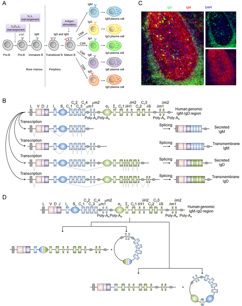
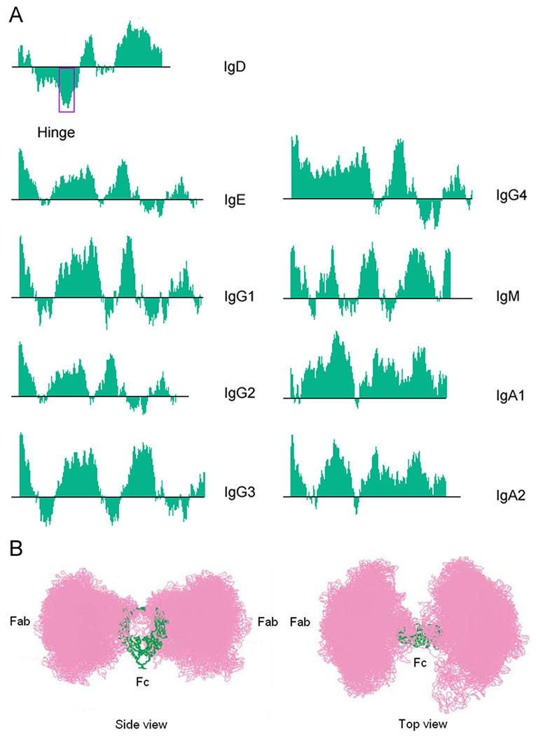
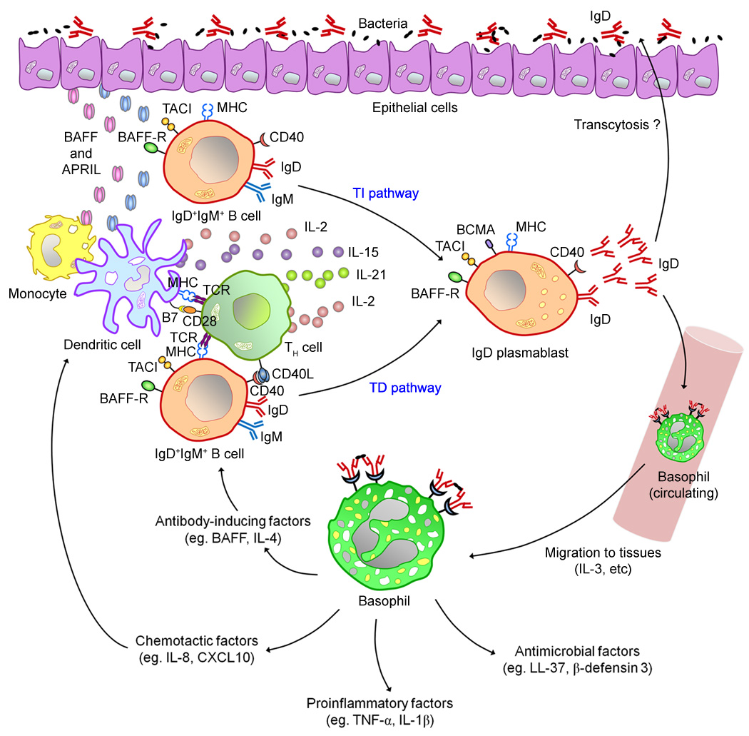
Immunoglobulin D (IgD) has remained a mysterious antibody class for almost half a century. IgD was initially thought to be a recently evolved Ig isotype expressed only by some mammalian species, but recent discoveries in fishes and amphibians demonstrate that IgD was present in the ancestor of all jawed vertebrates and has important immunological functions. The structure of IgD has been very dynamic throughout evolution. Mammals can express IgD through alternative splicing and class switch recombination. Active cell-dependent and T-cell-independent IgM-to-IgD class switching takes place in a unique subset of human B cells from the upper aerodigestive mucosa, which provides a layer of mucosal protection by interacting with many pathogens and their virulence factors. Circulating IgD can bind to myeloid cells such as basophils and induce antimicrobial, inflammatory, and B-cell-stimulating factors upon cross-linking, which contributes to not only immune surveillance but also inflammation and tissue damage when this pathway is overactivated under pathological conditions. Recent research shows that IgD is an important immunomodulator that orchestrates an ancestral surveillance system at the interface between immunity and inflammation.


Figures

Fig. 1
Fig. 1. Protein sequence alignment of mammalian IgD heavy chain hinge region
Sequences, with accession numbers in brackets, were downloaded from NCBI GenBank: human (P01880.2), chimpanzee (AAB89456.1), baboon (ABB89458.1), macaque (ABB89463.1), mangabey (ABB89466.1), pig (AAN03672.1), horse (AAU09794.1), cattle (AAN03673.1), sheep (AAN03671.1), dog (ABB89467.1), giant panda (AAX73311.1), mouse (P01881.2), and rat (P01883.1). Sequences were aligned using VectorNTI (Invitrogen Corporation). Hinge regions are divided into two sub-regions, with the second sub-region containing the conserved stretch of charged amino acids in primates. Hinge region boundary is based on the peptides encoded by the two hinge exons of human IgD.
Fig. 2
Fig. 2. Expression of human IgD by alternative splicing and CSR
(A) Ig isotype expression during human B-cell development. Pre-B cells and immature B cells developing in the bone marrow express μ heavy chain associated with either surrogate light chains VpreB and Vλ5 before light chain rearrangement or rearranged L chain after L chain rearrangement. Upon exiting the bone marrow, transitional and mature B cells co-express IgM and IgD through alternative splicing. IgD is expressed at a higher level on the cell surface than IgM. After antigenic stimulation, most mature B cells rapidly downregulate IgD expression and undergo class switch recombination (CSR) to express either IgG, IgA, or IgE in response to the antigenic signals encountered. Some human B cells also undergo CSR to IgD. Class-switched B cells can further differentiate into plasmablasts or plasma cells (PCs) that secrete the respective Ig isotype. Some mature B cells can downregulate IgD and develop into IgM-secreting PCs perhaps either without undergoing CSR or by undergoing ‘silent CSR’ involving intra-Sμ region DNA recombination (Chen and Cerutti, unpublished data). (B) Expression of IgD and IgM by alternative splicing in mature B cell. Exons encoding the rearranged VDJ region and the various domains (including the membrane and secreted portions) of IgM and IgD are shown in boxes. Dotted gray lines show the various splicing configurations of primary transcripts to yield secreted and transmembrane forms of IgM and IgD. (C) IgD and IgM expression in follicular mantle and germinal center B cells. A lymphoid follicle in human tonsil is shown. Mature B cells located in the follicular mantle zone prior to antigen encounter express more IgD (green) than IgM (red) on the surface due to the higher stability and translation efficiency of δ mRNA than μ mRNA. After antigenic stimulation, they enter the germinal center, where IgD is rapidly downregulated at transcriptional level and IgM is transcriptionally upregulated and the half-life of μ mRNA is significantly prolonged, as evidenced by the drastic reduction of IgD and increase in IgM in the germinal center. Cells that stain strongly with IgD in the germinal center are class switched IgD-secreting cells. Some IgM staining in the germinal center is contributed by follicular dendritic cells that capture IgM-containing immune complexes. DAPI (blue) stains cell nuclei. Original magnification, ×40. (D) Expression of IgD by CSR. Schematic representation of Cμ-to-Cδ CSR in human, which has been found to occur between Sμ and σδ regions (left) and between Iμ and Σμ regions (right) in normal and malignant B cells. The intervening DNA is looped out after CSR and forms a switch circle.
Fig. 3
Fig. 3. Human IgD hinge region contains a sequence resembling that of consensus ligand of heparin
(A) Hydrophobicity indices of human Ig isotypes at pH 3.4 determined in silico using the VectorNTI software. The region of high hydrophilicity in IgD is demarcated by a rectangle. Sequences of these proteins were downloaded from NCBI GenBank, with GenBank ID in brackets: IgD (P01880.2), IgE (P01854.1), IgG1 (AAH73782), IgG2 (AAH62335), IgG3 (AAH33178), IgG4 (AAh25985), IgM, (AAH89412), IgA1 (AAH87841), IgA2 (AAH73765). (B) Three-dimensional motions of the IgD Fab parts around the Fc part. The Fab parts are colored in pink and the Fc part is colored in green. The length and the semi-extended flexible conformation of the IgD hinge allow the Fab parts to swivel around the Fc part in three dimensions in solution. This motion gives the Fab parts extraordinary flexibility in capturing antigens, as well as renders the Fc part shielded by the Fab parts in solution. The Protein Data Bank (PDB) ID of the best-fit IgD structure predicted in this study is 1ZVO. Modified from (119) with permission. [Permission from Elsevier to use the figure shown in Fig. 3B, modified from (119), can be found at https://s100.copyright.com/CustomerAdmin/PLF.jsp?lID=2010031_1268678952456.]
Fig. 4
Fig. 4. Model of human IgD regulation and function
B cells from the upper aerodigestive mucosa undergo CSR from Cμ to Cδ through local T-dependent (TD) and T-indepdent (TI) pathways involving CD40L, BAFF, or APRIL together with a unique cocktail of cytokines, including IL-2 and IL-21 or IL-15 and IL-21. CSR requires AID and generates mucosal and circulating IgD plasmablasts that secrete IgD reactive against respiratory bacteria. Mucosal IgD might enhance local immunity after translocating across epithelial cells, whereas circulating IgD binds to basophils. In addition to delivering rapid innate immune signals and alerting the immune system as to the presence of invading bacteria, basophils exposed to IgD-reactive antigens migrate to systemic and mucosal lymphoid organs, perhaps in response to chemotactic factors released by IgD-stimulated mast cells. Tissue-based basophils enhance immune protection by releasing antimicrobial, opsonizing, B-cell-stimulating, chemotactic and pro-inflammatory mediators such as cathelicidin, IL-1β, IL-4, IL-8, IL-13, CD40L, BAFF, APRIL, TNF, and CXCL10, but not histamine. Finally, IgD-armed basophils may regulate B-cell homeostasis through tonic release of the obligatory B cell survival factor BAFF in response to IgD-reactive foreign or autologous antigens.

References

    1. Rowe DS, Fahey JL. A new class of human immunoglobulins. I. A unique myeloma protein. J Exp Med. 1965;121:171–184. - PMC - PubMed
    1. Rowe DS, Fahey JL. A new class of human immunoglobulins. Ii. Normal serum IgD. J Exp Med. 1965;121:185–199. - PMC - PubMed
    1. Black CA. A brief history of the discovery of the immunoglobulins and the origin of the modern immunoglobulin nomenclature. Immunol Cell Biol. 1997;75:65–68. - PubMed
    1. Finkelman FD, van Boxel JA, Asofsky R, Paul WE. Cell membrane IgD: demonstration of IgD on human lymphocytes by enzyme-catalyzed iodination and comparison with cell surface Ig of mouse, guinea pig, and rabbit. J Immunol. 1976;116:1173–1181. - PubMed
    1. Martin LN, Leslie GA, Hindes R. Lymphocyte surface IgD and IgM in non-human primates. Int Arch Allergy Appl Immunol. 1976;51:320–329. - PubMed

Publication types