. 2010 Aug 20:10:89.
doi: 10.1186/1471-213X-10-89.

Pax2 and Pax8 cooperate in mouse inner ear morphogenesis and innervation

Affiliations

Pax2 and Pax8 cooperate in mouse inner ear morphogenesis and innervation

Maxime Bouchard et al. BMC Dev Biol. .

Abstract

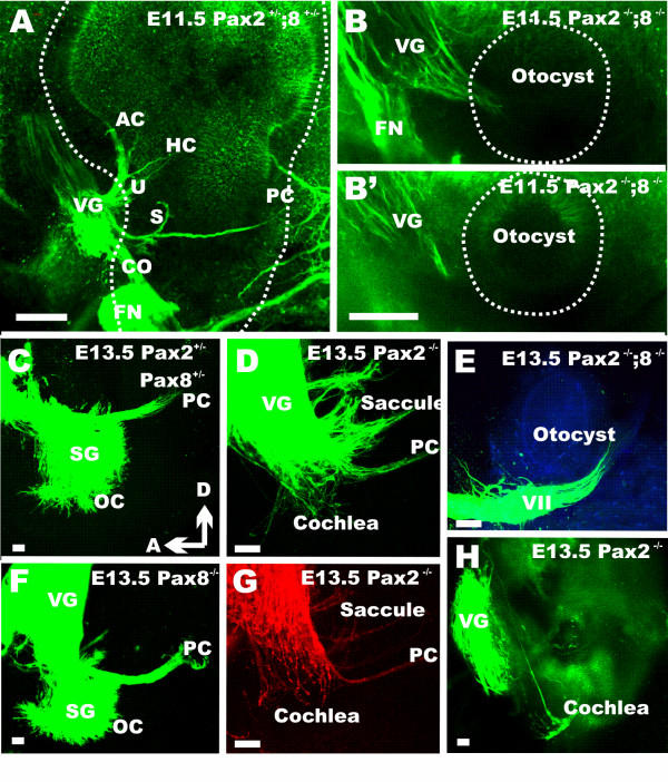
Background: Pax2;5;8 transcription factors play diverse roles in vertebrate and invertebrate organogenesis, including the development of the inner ear. Past research has suggested various cochlear defects and some vestibular defects in Pax2 null mice but the details of the cochlear defects and the interaction with other Pax family members in ear development remain unclear.

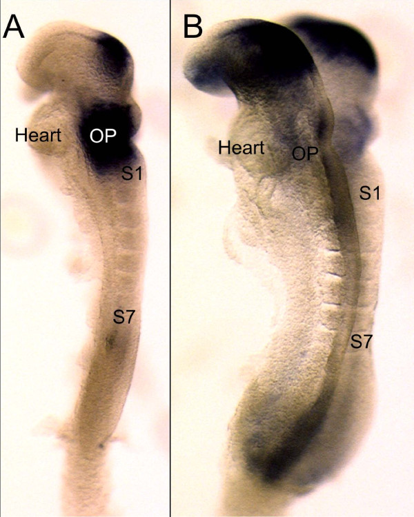
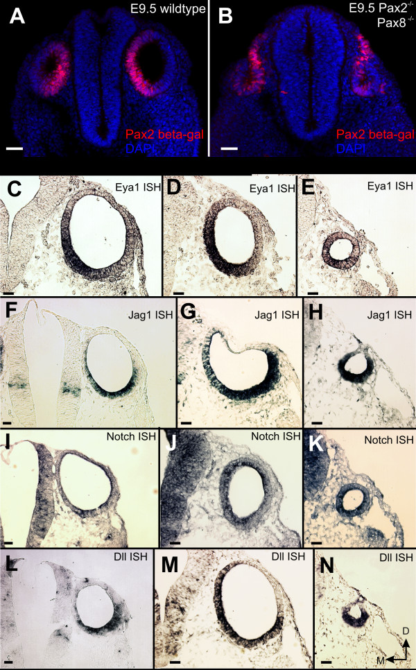
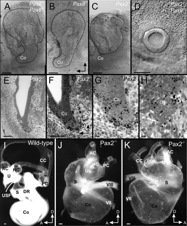
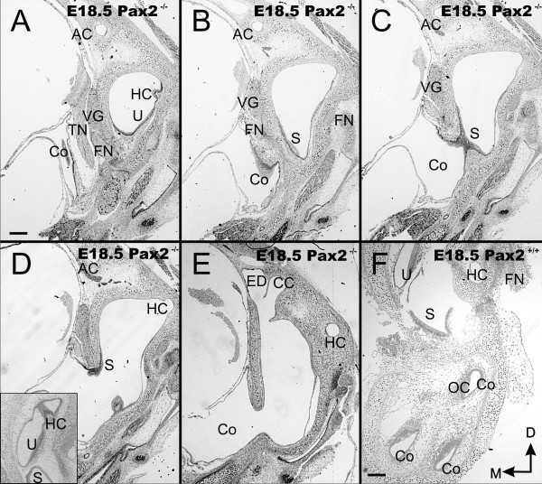
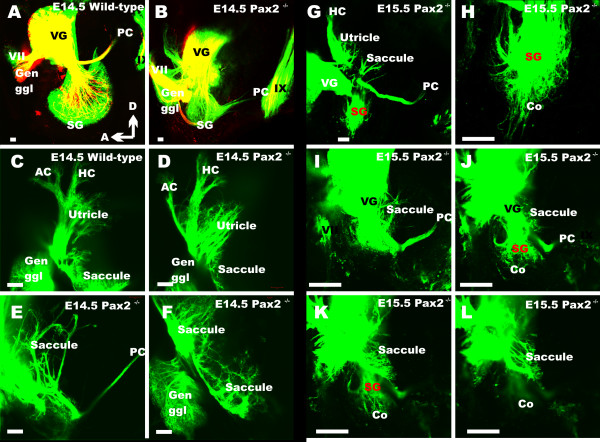
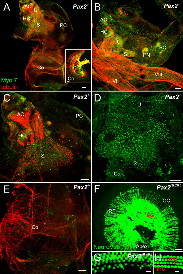
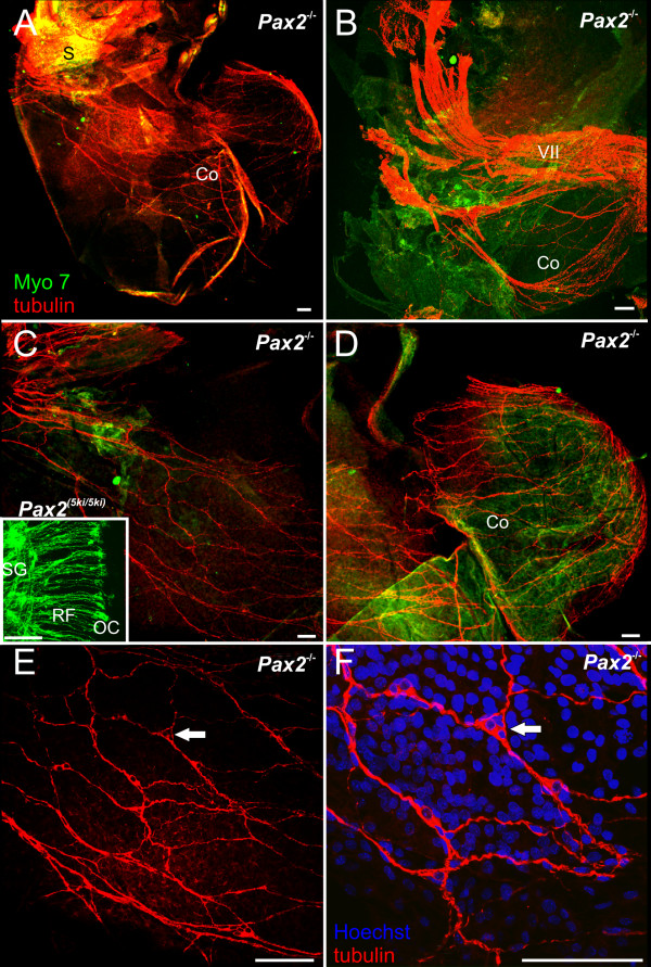
Results: We show that Pax2;8 double null mice do not develop an ear past the otocyst stage and show little to no sensory as well as limited and transient neuronal development, thus indicating that these two family members are essential for overall ear morphogenesis and sustained neurosensory development. In support of functional redundancy between Pax proteins, Pax2 can be substituted by a Pax5 minigene, a gene normally not expressed in the embryonic mouse ear. There is no detectable morphological defect in Pax8 null mice suggesting that Pax2 expression can compensate for Pax8. Conversely, Pax8 cannot compensate for Pax2 leading to a cochlear phenotype not fully appreciated previously: Cochlear development is delayed until E15.5 when the cochlea extrudes as a large sack into the brain case. Immunocytochemistry and tracing from the brain show that a cochlear spiral ganglia form as a small addition to the inferior vestibular ganglion. However, the empty cochlear sack, devoid of any sensory epithelium development as indicated by the absence of Sox2 or MyoVII expression, nevertheless develop a dense innervation network of small neurons situated in the wall of the cochlear sack.

Conclusions: Combined these data suggest that Pax2 is needed for organ of Corti formation and is directly or indirectly involved in the coordination of spiral ganglion formation which is partially disrupted in the Pax2 null ears. All three Pax genes can signal redundantly in the ear with their function being determined primarily by the spatio-temporal expression driven by the three distinct promoters of these genes.


Figures

Figure 1
Figure 1
Early expression of Pax2 and Pax8 compared. This image shows the earliest expression of Pax8 (A) and Pax2 (B) in 7 somite (S1, S7) mouse embryos of approximately 8 embryonic days. Note that at this stage Pax8 is more profoundly expressed throughout the area of the future otic placode (OP) just anterior to the first somite (S1). In contrast, Pax2 is more prominently expressed in the brain with only very limited expression in the area of the otic placode (OP). At this stage the heart is anterior to the future otic placode. Bar indicates 100 um.
Figure 2
Figure 2
Pax2;8 affects invagination but not early marker expression. Shortly after the otocyst is detached from the ectoderm, Pax2 expression is throughout the ventral and medial aspect of the developing ear (as revealed by a LacZ-reporter for Pax2). In contrast, the reporter for Pax2 shows in the Pax2;8 double null mouse a much wider expression including areas outside the partially invaginated otic placode. Expression profile of four genes known to be important for overall ear development (Eya1, Dll) and for neurosensory development (Jag1, Notch) show no expression changes in simple Pax2 or compound Pax2;8 double mutants, suggesting that Pax2 and Pax8 have little effect on these early markers. However, there is a reduction in otocyst size and a delayed detachment from the ectoderm in Pax2;8 double null mice. Bar indicates 100 um.
Figure 3
Figure 3
Overview of ear defects and sections. Whole mounted (A-D, I-K) and coronally sectioned ears (E-H) show the mutant effects on morphology. The cochlear duct is truncated in Pax2 null whereas Pax2;8 double null mice have only an otic vesicle at E11.5 (A, B; outlined by a dotted line). Coronal sections (see vertical line in C) show no cochlear outgrowth in Pax2 null mice as late as E13.5 (E). Numerous apoptotic cells in and around the cochlear duct (F, G) are indicated by chromatin condensation. Cartilage is obvious lateral but not medial to the ear (E). Higher magnification shows also apoptotic cells in the ganglion including in the more ventral, presumably spiral ganglion part (H). At E18.5 wild-type ears have developed a coiled cochlea, three distinct canals and recesses for the saccule and the utricle (I). The utricle is separated by the utriculo-saccular foramen from the saccule (USF in I) and the cochlea is separated from the saccule by the ductus reunions (DR in I). Pax2 null mice have an inflated saccule and cochlear duct with shortened semicircular canals (J, K). The only constriction resulting in two adjacent vesicles is near the saccule. Facial (VII) and vestibular (VIII) nerves are abutting the ear adjacent to the constriction separating the superior from the inferior division. AC, anterior canal/crista; CC, common crus; HC, horizontal canal/crista; Co, cochlear duct/sack; DR, ductus reunions; Ggl, ganglion; PC, posterior canal/crista; S, Saccule; U, utricle; USF, utriculo-saccular foramen; VII, facial nerve; VIII vestibular/cochlear nerve. Bar indicates 100 um in A-F, I-K, 10 um in G, L.
Figure 4
Figure 4
Overview of sections through E18.5 ear. These near serial coronal sections through the ear show the unusual morphology with a vesicle within the otic capsule (top of each section) and the large ventral vesicle that is expanded into the brain cavity, labeled as cochlea (C in A-E) instead of the multiple cross sections through the cochlear duct found in wild-type (F). Note the brain has been removed to verify before sectioning that a vesicle was present. Canals and canal cristae can be found along the superior vesicle surrounded by periodic mesenchyme. However, the horizontal crista is not in the horizontal canal (HC in A, D) whereas it sits in wild-type at the orifice of the horizontal canal (insert in D). Hair cells of the utircular and saccular macula are continuous and extend into the ventral, extruded sack of the ear (A-D). While adjacent to each other, the endolymphatic duct (ED) and the common crus (CC) are nevertheless distinct anatomical entities (E). AC, anterior canal/crista; CC, common crus; FN, facial nerve; ED, endolymphatic duct; HC, horizontal canal/crista; Co, cochlea duct/sack; OC, organ of Corti; PC, posterior canal/crista; S, saccule; TN, trigeminal nerve; U, utricle; VG, vestibular ganglion. Bar indicates 100 um.
Figure 5
Figure 5
The pattern of innervations at E11.5 and E13.5. Innervation of the ear in terms of distinct fibers to sensory epithelia develops around E11.5 in wild-type mice (A). In Pax2;8 double null mutants there is at least a transient projection to a small otocyst which never develops any distinct projection pattern to the otocyst. At later stages, Pax2;8 double null mice have only an ear vesicle without any feature that is completely devoid of any afferent or efferent innervation (E, compare to C, D). In contrast, Pax2; Pax8 double heterozygote (C) and Pax8 null mouse (F) show at E13.5 fibers reaching to the posterior crista (PC) and the spiral ganglia (Sg) extend some fibers to the developing organ of Corti (OC). In contrast, fibers to the PC are reduced in Pax2 null mice, the saccular innervation is diffuse and there is little extension of afferent (D) or efferent (G) nerve fibers to the cochlea. Note the similarities in distribution of afferents (D) and efferents (G) in these two Pax2 null mice indicating a consistent but different pattern of innervation for these mutants. Injection of dye selectively into the otocyst reveals a few fibers extending toward the brain from the region of this presumptive cochlea (H). These data suggest that at least some early development of the cochlea neurons occurs but that the overall development or projection to the brain is severely truncated. OC, organ of Corti; PC, posterior crista; SG, spiral ganglion; VG, vestibular ganglion; VII, facial nerve, Bar indicates 100 um.
Figure 6
Figure 6
The pattern of innervation at E14.5 and E15.5. Wild-type animals show afferents (in green) to the growing cochlear spiral (A). Efferent fibers, shown in red, begin to form the intraganglionic spiral bundle. There is no cochlear spiral or spiral ganglion in Pax2 null mice (E). Instead there is an aggregation of neurons near the reduced fibers leading to the posterior canal crista (B), the presumed spiral ganglion (SG). In mutants, efferent fibers reroute from the spiral ganglion to the facial nerve (B). The innervation to the anterior canal crista (AC in C, D) is reduced, but not that to the horizontal crista (C, D). The saccule shows near normal organization of fibers in the sensory epithelium (F) as well as an overshooting projection at a different focal plane (E). At E15.5, sensory neurons that can be filled with brain injections of lipophilic dyes form a tear drop arrangement of spiral sensory neurons (G, H). Multiple nerve fibers radiate away for a short distance toward the cochlear expansion (CO), but no pattern reminiscent of radial fibers in the cochlea can be observed (G, H, J, K). There is a variable reduction of nerve fibers to the anterior and posterior canal crista (D, G) and the innervation of the saccule is variable (G-L). A focal series (I-L) shows that spiral ganglion neurons are distinct in their distribution from vestibular ganglion cells as is evidenced by. AC, anterior canal crista; HC, horizontal canal crista; Co, cochlear duct/sack; Gen Ggl, geniculate ganglion; PC, posterior canal crista; VG, vestibular ganglion; IX, glossopharyngeal nerve. Bar indicates 100 um.
Figure 7
Figure 7
Vestibular hair cells and some innervation develop in Pax2 null mice. Pattern of innervations and distribution of hair cells as revealed by anti-tubulin (red) and anti-Myo 7 (green) antibodies (A-E, G, H) and lipophilic dye tracing (insert in A, F). Hair cells are concentrated to the superior part of the ear in Pax2 null mice (A, C, D), innervations exists to vestibular (A-C) and cochlear sack (A, E). Pax2 5ki/5ki show a near normal innervation of the cochlea (F) compared to that found in wild-type (insert in A). Anterior, horizontal and posterior canal and crista can be recognized in all ears (compare C, F) and may receive no or few nerve fibers. Myosin 7 labeled hair cells form an unstructured aggregation tentatively divided into utricle (U) saccule (S). In addition to several small isolated patches of hair cells we found aeas without hair cells near the ventral border of the saccular patch (A-D). Innervation implies that nerve fiber pathfinding is unaltered. The cochlear sack has no hair cells and a diffuse innervation. This contrasts to the radial fibers (RF) to the inner and outer hair cells (IHC, OHC) of the organ of Corti (OC) with the in Pax2 5ki/5ki (F, G) and wild-type cochleae (insert in A, H). AC, anterior canal crista; HC, horizontal canal crista; IHC, inner hair cell; OC, organ of Corti; OHC, outer hair cell; PC, posterior canal crista; RF, radial fibers; S, saccule; SG, spiral ganglion; U, utricle. Bar indicates 100 um.
Figure 8
Figure 8
A cochlear sack innervation develops as dispersed neurons in Pax2 null mice. Details of the innervation of the cochlear sack are shown using anti-tubulin immoncytochemistry in Pax2 null mice (red, A-F) and lipophilic tracing in wild-type mice (insert in C). In contrast to the patterned innervation of the organ of Corti in wild-type mice by radial fibers emanating from spiral ganglia (insert in C) innervation of the cochlear sack of the mutant is provided by an irregular network of nerve fibers (A, C, D). A continuity of this network with the vestibular fibers (B) could not be verified by tracing from the brain, suggesting that even the fibers that extent toward the saccule may not project to the brain for labeling. Higher magnification (E, F) shows a network of neurons that appears to be interconnected by fibers thus generating the network that is so prominent at lower magnification throughout the cochlear sack. Bar indicates 100 um.

References

    1. Srivastava M, Simakov O, Chapman J, Fahey B, Gauthier ME, Mitros T, Richards GS, Conaco C, Dacre M, Hellsten U. et al.The Amphimedon queenslandica genome and the evolution of animal complexity. Nature. 2010;466(7307):720–726. doi: 10.1038/nature09201. - DOI - PMC - PubMed
    1. Kozmik Z, Daube M, Frei E, Norman B, Kos L, Dishaw LJ, Noll M, Piatigorsky J. Role of Pax genes in eye evolution: a cnidarian PaxB gene uniting Pax2 and Pax6 functions. Dev Cell. 2003;5(5):773–785. doi: 10.1016/S1534-5807(03)00325-3. - DOI - PubMed
    1. Burton Q, Cole LK, Mulheisen M, Chang W, Wu DK. The role of Pax2 in mouse inner ear development. Dev Biol. 2004;272(1):161–175. doi: 10.1016/j.ydbio.2004.04.024. - DOI - PubMed
    1. Favor J, Sandulache R, Neuhauser-Klaus A, Pretsch W, Chatterjee B, Senft E, Wurst W, Blanquet V, Grimes P, Sporle R. et al.The mouse Pax2(1Neu) mutation is identical to a human PAX2 mutation in a family with renal-coloboma syndrome and results in developmental defects of the brain, ear, eye, and kidney. Proc Natl Acad Sci USA. 1996;93(24):13870–13875. doi: 10.1073/pnas.93.24.13870. - DOI - PMC - PubMed
    1. Torres M, Gomez-Pardo E, Gruss P. Pax2 contributes to inner ear patterning and optic nerve trajectory. Development. 1996;122(11):3381–3391. - PubMed

Publication types

MeSH terms