. 2010 Aug 20:9:220.
doi: 10.1186/1476-4598-9-220.

Nicotine-induced survival signaling in lung cancer cells is dependent on their p53 status while its down-regulation by curcumin is independent

Affiliations

Nicotine-induced survival signaling in lung cancer cells is dependent on their p53 status while its down-regulation by curcumin is independent

Vineshkumar T Puliyappadamba et al. Mol Cancer. .

Abstract

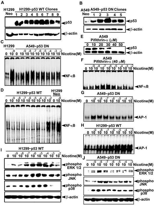
Background: Lung cancer is the most lethal cancer and almost 90% of lung cancer is due to cigarette smoking. Even though nicotine, one of the major ingredients of cigarette smoke and the causative agent for addiction, is not a carcinogen by itself, several investigators have shown that nicotine can induce cell proliferation and angiogenesis. We observed that the proliferative index of nicotine is different in the lung cancer cell lines H1299 (p53-/-) and A549 (p53+/+) which indicates that the mode of up-regulation of survival signals by nicotine might be different in cells with and without p53.

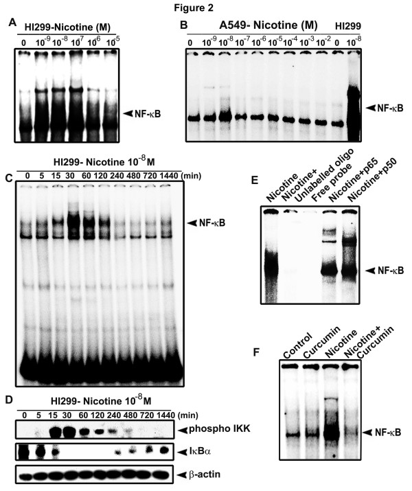
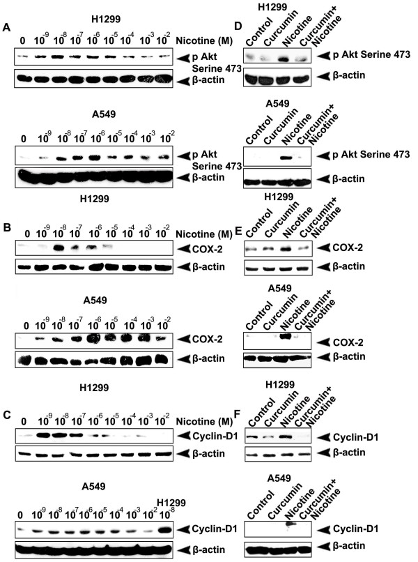
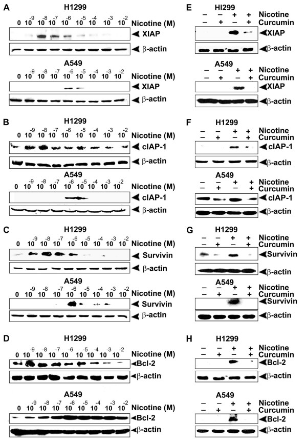
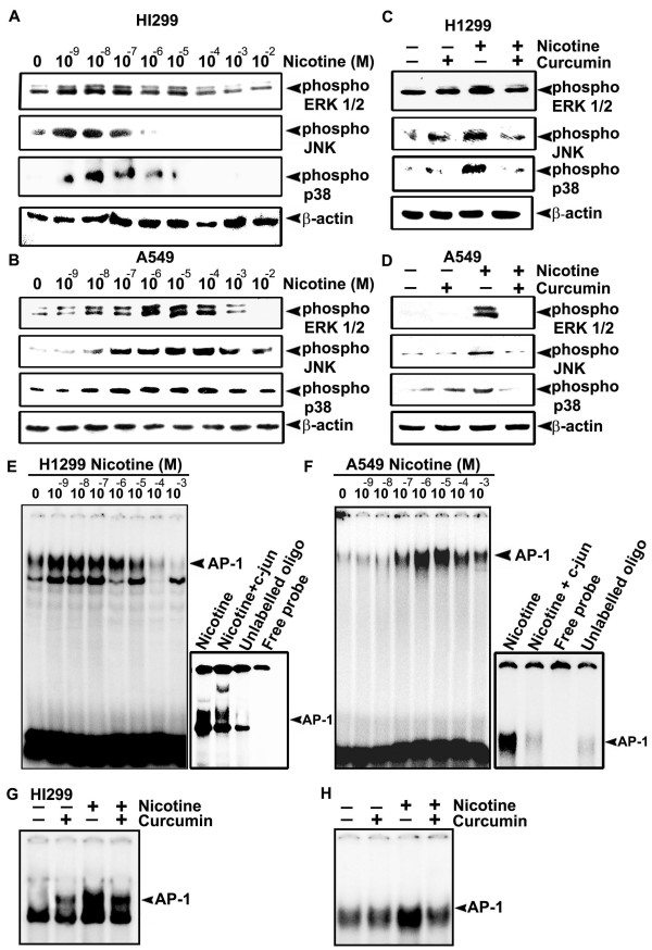
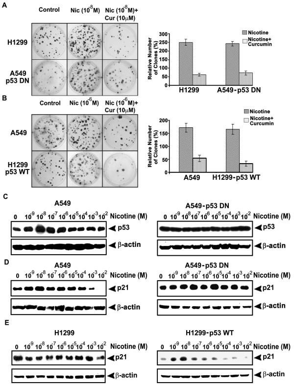
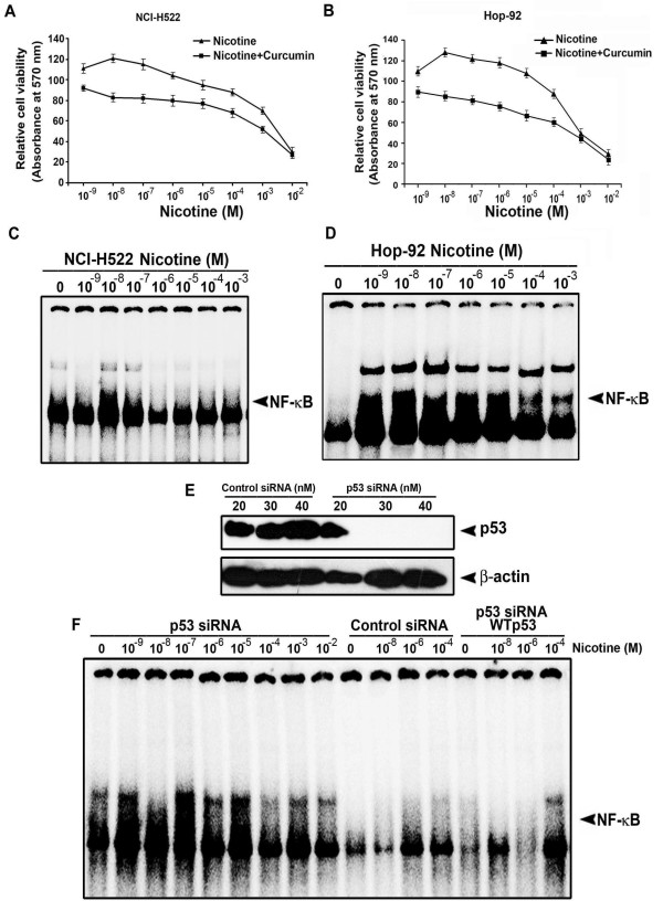
Results: While low concentrations of nicotine induced activation of NF-κB, Akt, Bcl2, MAPKs, AP1 and IAPs in H1299, it failed to induce NF-κB in A549, and compared to H1299, almost 100 times higher concentration of nicotine was required to induce all other survival signals in A549. Transfection of WT-p53 and DN-p53 in H1299 and A549 respectively, reversed the mode of activation of survival signals. Curcumin down-regulated all the survival signals induced by nicotine in both the cells, irrespective of their p53 status. The hypothesis was confirmed when lower concentrations of nicotine induced NF-κB in two more lung cancer cells, Hop-92 and NCI-H522 with mutant p53 status. Silencing of p53 in A549 using siRNA made the cells susceptible to nicotine-induced NF-κB nuclear translocation as in A549 DN-p53 cells.

Conclusions: The present study reveals a detrimental role of nicotine especially in lung cancer patients with impaired p53 status and identifies curcumin as a potential chemopreventive.


Figures

Figure 1
Figure 1
Nicotine induces more proliferation in H1299 than in A549, which is down regulated by curcumin. (A-B) The cells were treated with nicotine and/or curcumin for 72 h and 24 h respectively for MTT assay and thymidine incorporation assay. Tritiated thymidine (0.5 μCi/well) was added 6 h before the completion of incubation. All error bars indicate standard deviation between three independent experiments.
Figure 2
Figure 2
Nicotine activates NF-κB in H1299, but not in A549. (A-B) Nuclear extracts were prepared from the cells treated with indicated concentrations of nicotine for 30 min and NF-κB nuclear translocation was assessed by EMSA. (C) H1299 cells were treated with 10-8 M nicotine for different time intervals and EMSA was done. (D) The cytosolic extracts of the time kinetic study of NF-κB activation were Western blotted against the respective antibodies. (E) The nuclear extract of nicotine-treated H1299 cells were incubated with p50 and p65 antibodies or unlabelled oligo and EMSA was conducted to detect supershift and cold competition respectively. (F) H1299 cells were treated with curcumin and then with nicotine for 30 min and EMSA was done. All blots and EMSAs are representative samples of three independent experiments.
Figure 3
Figure 3
Curcumin inhibits nicotine-induced phosphorylation of Akt and over expression of COX-2 and Cyclin D1. (A-C) The cells were treated with various concentrations of nicotine (30 min for phospho-Akt and 24 h for COX-2 and Cyclin-D1) and whole cell lysates prepared were blotted against phospho-Akt, COX-2 and Cyclin-D1 antibodies. (D-F) The cells were treated with curcumin and then with nicotine (10-8 M in H1299 and 10-6 M in A549) and whole cell lysates were blotted as in (A-C). All blots are representative samples of three independent experiments.
Figure 4
Figure 4
Nicotine-induced over expression of IAPs and Bcl2 is inhibited by curcumin. (A-D) The cells were treated with nicotine for 24 h and whole cell lysates were Western blotted using antibodies against XIAP, cIAP-1 survivin and Bcl2. (E-H) The cells were treated with curcumin and then with nicotine (10-8M for H1299 and 10-6 M for A549) and whole cell lysates were blotted as in (A-D). All blots are representative samples of three independent experiments.
Figure 5
Figure 5
Curcumin downregulates nicotine-induced phosphorylation of MAPKs and nuclear translocation of AP-1. (A-B) The cells were treated with nicotine (10-9-10-2M) for 30 min and whole cell lysates were blotted against respective phospho specific antibodies. (C-D) The cells were treated with curcumin and then with nicotine (10-8M for H1299 and 10-6 M for A549) and whole cell lysates were blotted as in (A-B). (E-F) Nuclear extracts were prepared from the cells treated with nicotine (10-9-10-2M) for 30 min. and AP-1 activation was assessed by EMSA. Super shift and cold competition was done by incubating the nuclear extract with c-jun antibody and unlabelled oligo respectively. (G-H) The cells were treated with curcumin and then with nicotine (10-8M for H1299 and 10-6 M for A549) and EMSA was done using nuclear extracts. All blots and EMSAs are representative samples of three independent experiments.
Figure 6
Figure 6
Over-expression and silencing of p53 produce reciprocal effect on the activation pattern of NF-κB, MAPKs and AP-1. (A-B) H1299 and A549 cells were transfected with pcDNA3-p53 WT and pcDNA3-p53 DN constructs respectively and the stable clones selected were lysed and Western blotted against p53 antibody. (C-D) H1299-p53 WT and A549-p53 DN cells were treated with nicotine (10-9-10-2M) for 30 min, and EMSA was done using nuclear extracts. (E) A549 cells were treated with different concentrations of pifithrin-α (10-50 μM) for 4 h and the whole cell lyasates prepared were Western blotted against p53 antibody. (F) A549 cells were treated with 40 μM pifithrin-α for 4 h followed by nicotine (10-9-10-2M) for 30 min and EMSA was done. (G-H) H1299-p53 WT and A549-p53 DN cells were treated with nicotine (10-9-10-2M) for 30 min, and EMSA was done. (I-J) H1299 WT-p53 and A549 DN-p53 cells were treated with nicotine (10-9-10-2M) for 30 min and whole cell lysates were Western blotted against respective phospho specific antibodies. All blots and EMSAs are representative samples of three independent experiments.
Figure 7
Figure 7
Curcumin inhibits enhancement of clonogenicity by nicotine, which induces p53 and p21 in A549 and p21 in H1299-p53 WT. (A-B) The cells were treated with nicotine and/or curcumin for 72 h, after which cells were split and seeded in six well plates, kept for 2 weeks and clones developed were stained and counted. (C-E) The cells were treated with nicotine (10-9-10-2M) for 24 h and whole cell lysates were Western blotted against p53 antibody. All figures are representative samples of three independent experiments.
Figure 8
Figure 8
Nicotine induces proliferation and NF-κB nuclear translocation in Hop-92, NCI-H522 and p53-silenced A549 cells. (A-B) The cells were treated with nicotine and/or curcumin and cell viability was measured by MTT assay. All error bars indicate standard deviation between three independent experiments. (C-D) Nuclear extracts were prepared from the cells treated with indicated concentrations of nicotine for 30 min and NF-κB nuclear translocation was assessed by EMSA. (E) A549 cells were transfected with indicated concentrations of control-si RNA or p53-si RNA and after 48 h, the whole cell lysates were Western blotted against p53 antibody. (F) A549 cells were transfected with indicated concentrations of control-si RNA or p53-si RNA and after 48 h, treated with indicated concentrations of nicotine for 30 min and NF-κB nuclear translocation was assessed by EMSA. All blots and EMSAs are representative samples of three independent experiments.

References

    1. D'Amico D, Carbone D, Mitsudomi T, Nau M, Fedorko J, Russell E, Johnson B, Buchhagen D, Bodner S, Phelps R, Gazdar A, Minna JD. High frequency of somatically acquired p53 mutations in small-cell lung cancer cell lines and tumors. Oncogene. 1992;7:339–346. - PubMed
    1. Waldum HL, Nilsen OG, Nilsen T, Rørvik H, Syversen V, Sanvik AK, Haugen OA, Torp SH, Brenna E. Long-term effects of inhaled nicotine. Life Sciences. 1996;58:1339–1346. doi: 10.1016/0024-3205(96)00100-2. - DOI - PubMed
    1. Heeschen C, Weis M, Aicher A, Dimmeler S, Cooke JP. A novel angiogenic pathway mediated by non-neuronal nicotinic acetylcholine receptors. J Clin Inves. 2002;110:527–536. - PMC - PubMed
    1. Dasgupta P, Rastogi S, Pillai S, Ordonez-Ercan D, Morris M, Haura E, Chellappan S. Nicotine induces cell proliferation by β-arrestin-mediated activation of Src and Rb-Raf-1 pathways. J Clin Invest. 2006;116:2208–2217. doi: 10.1172/JCI28164. - DOI - PMC - PubMed
    1. Egleton RD, Brown KC, Dasgupta P. Angiogenic activity of nicotinic acetylcholine receptors: Implications in tobacco related vascular diseases. Pharmacol Ther. 2009;121:205–223. doi: 10.1016/j.pharmthera.2008.10.007. - DOI - PubMed

MeSH terms