. 2010 Sep 10;87(3):365-70.
doi: 10.1016/j.ajhg.2010.07.020. Epub 2010 Aug 19.

TBC1D24, an ARF6-interacting protein, is mutated in familial infantile myoclonic epilepsy

Affiliations

TBC1D24, an ARF6-interacting protein, is mutated in familial infantile myoclonic epilepsy

Antonio Falace et al. Am J Hum Genet. .

Abstract

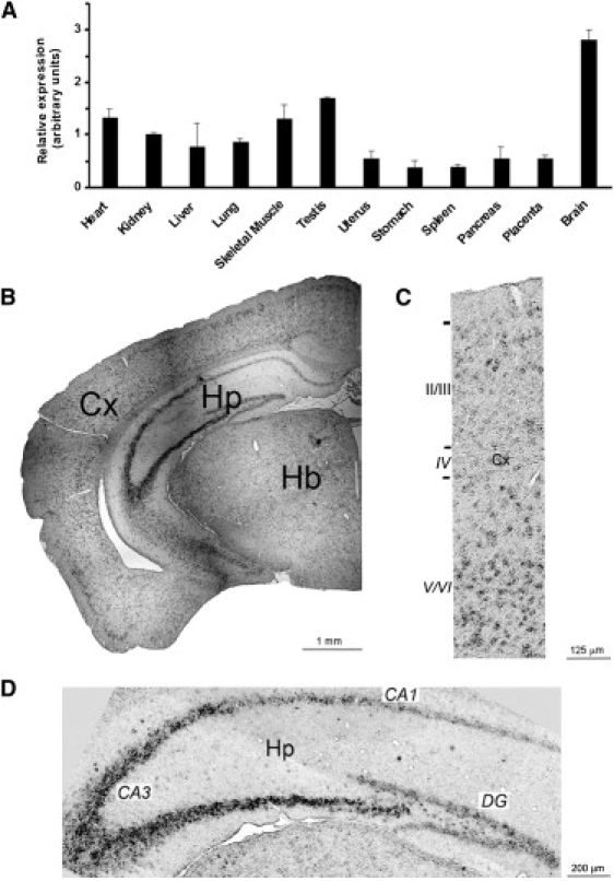
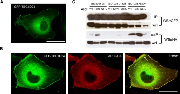
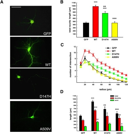
Idiopathic epilepsies (IEs) are a group of disorders characterized by recurrent seizures in the absence of detectable brain lesions or metabolic abnormalities. IEs include common disorders with a complex mode of inheritance and rare Mendelian traits suggesting the occurrence of several alleles with variable penetrance. We previously described a large family with a recessive form of idiopathic epilepsy, named familial infantile myoclonic epilepsy (FIME), and mapped the disease locus on chromosome 16p13.3 by linkage analysis. In the present study, we found that two compound heterozygous missense mutations (D147H and A509V) in TBC1D24, a gene of unknown function, are responsible for FIME. In situ hybridization analysis revealed that Tbc1d24 is mainly expressed at the level of the cerebral cortex and the hippocampus. By coimmunoprecipitation assay we found that TBC1D24 binds ARF6, a Ras-related family of small GTPases regulating exo-endocytosis dynamics. The main recognized function of ARF6 in the nervous system is the regulation of dendritic branching, spine formation, and axonal extension. TBC1D24 overexpression resulted in a significant increase in neurite length and arborization and the FIME mutations significantly reverted this phenotype. In this study we identified a gene mutation involved in autosomal-recessive idiopathic epilepsy, unveiled the involvement of ARF6-dependent molecular pathway in brain hyperexcitability and seizures, and confirmed the emerging role of subtle cytoarchitectural alterations in the etiology of this group of common epileptic disorders.


Figures

Figure 1
Figure 1
Genetic Analysis of TBC1D24 (A) Pedigree of the family with FIME and segregation analysis of TBC1D24 mutations. Haplotypes are shown in colored bars. The red and green haplotypes cosegregate with c.1526C>T and c.439G>C TBC1D24 mutations, respectively. Affected patients are indicated by filled symbols. (B) Electropherograms of c.439G>C and c.1526C>T mutations. (C) Genomic organization and functional domains of human TBC1D24. Affected amino acids are highly conserved throughout evolution.
Figure 2
Figure 2
Expression Analysis of TBC1D24 (A) RT-PCR showing the expression profile of TBC1D24 in various human tissues. TBC1D24 is preferentially expressed in brain. Data are represented as means ± SEM. (B) RNA in situ hybridization on brain sections from 12-week-old mouse shows that Tbc1d24 is abundantly expressed in the neocortex (Cx) and hippocampus (Hp). (C) High-magnification image of the cerebral cortex revealed a higher expression of Tbc1d24 in deep cortical layers (V/VI) compared to superficial layers (II-III). (D) High-magnification image of hippocampus shows the highest expression of Tbc1d24 in the CA3 region compared to the CA1 region and dentate gyrus (DG). Hb, hindbrain.
Figure 3
Figure 3
TBC1D24 Is an ARF6-Interacting Protein (A) Representative image of a COS-7 cell transfected with GFP-TBC1D24 and visualized 36 hr after transfection. (B) Representative images of a COS-7 cell cotransfected with GFP-TBC1D24 and ARF6-HA and visualized 36 hr after transfection. HA immunoreactivity was detected by HA antibody and Alexa546 conjugated secondary antibodies. The arrow in the merge panel shows colocalization of overexpressed ARF6 and TBC1D24 at the plasma membrane. Scale bars represent 20 μm. (C) TBC1D24-WT, D147H, and A509V were cotransfected with HA-tagged wild-type (WT), T27N, or Q67L ARF6 in COS-7 cells and immunoprecipitated (IP) with GFP antibody. Associated ARF6 (coIP) was detected by HA antibody (αHA) in retrospective western blotting (WB). Wcl, whole cell lysate.
Figure 4
Figure 4
Functional Analysis of WT and Mutant TBC1D24 (A) Representative images of mouse cortical neurons transfected at 6 DIV with either GFP, GFP-TBC1D24-wt (WT), GFP-TBC1D24-D147H (D147H), or GFP-TBC1D24-A509V (A509V) and visualized 36 hr after transfection. Scale bar represents 50 μm. (B and C) Overexpression of TBC1D24 provoked a massive increase in neurite length (B) and branching (C), whereas overexpression of its epileptogenic mutants (i.e., D147H and A509V) reverted this phenotype. (D) Quantitative analysis of neurite (total), dendrite, or axon length in 33 cells for each genotype. Dendrites and axons were distinguished by MAP2 labeling and/or morphology. Data expressed as means ± SEM and compared via one-way analysis of variance (ANOVA, B and C) or repeated-measures ANOVA (D) followed by the Bonferroni's multiple comparison test. ∗∗∗p < 0.0001, ∗∗p < 0.001 versus GFP; °°°p < 0.0001, °°p < 0.001, °p < 0.01 versus GFP-TBC1D24-wt.

References

    1. Hauser W.A., Annegers J.F., Kurland L.T. Incidence of epilepsy and unprovoked seizures in Rochester, Minnesota: 1935-1984. Epilepsia. 1993;34:453–468. - PubMed
    1. Turnbull J., Lohi H., Kearny J.A., Rouleau G.A., Delgado-Escueta M.H., Cossette P., Minassian B.A. Sacred disease secrets revealed: The genetics of human epilepsy. Hum. Mol. Genet. 2005;14:2491–2500. - PubMed
    1. Avanzini G., Franceschetti S. Cellular biology of epileptogenesis. Lancet Neurol. 2003;2:33–42. - PubMed
    1. Zhou Y.D., Lee S., Jin Z., Wright M., Smith S.E., Anderson M.P. Arrested maturation of excitatory synapses in autosomal dominant lateral temporal lobe epilepsy. Nat. Med. 2009;15:1208–1214. - PMC - PubMed
    1. de Nijs L., Léon C., Nguyen L., Loturco J.J., Delgado-Escueta A.V., Grisar T., Lakaye B. EFHC1 interacts with microtubules to regulate cell division and cortical development. Nat. Neurosci. 2009;12:1266–1274. - PubMed

Publication types

MeSH terms