. 2010 Sep 3;142(5):749-61.
doi: 10.1016/j.cell.2010.07.040. Epub 2010 Aug 19.

Structural basis of semaphorin-plexin recognition and viral mimicry from Sema7A and A39R complexes with PlexinC1

Affiliations

Structural basis of semaphorin-plexin recognition and viral mimicry from Sema7A and A39R complexes with PlexinC1

Heli Liu et al. Cell. .

Abstract

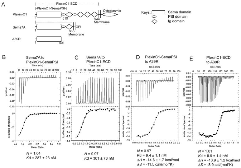
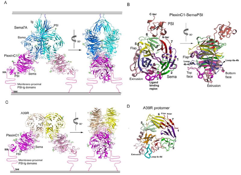
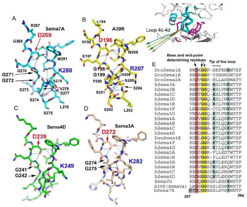
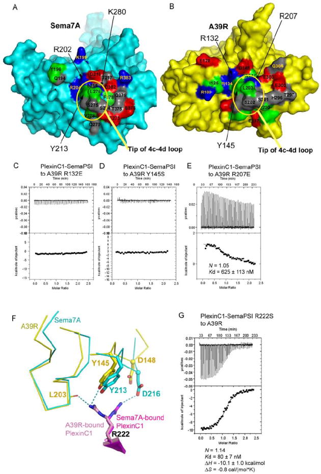
Repulsive signaling by Semaphorins and Plexins is crucial for the development and homeostasis of the nervous, immune, and cardiovascular systems. Sema7A acts as both an immune and a neural Semaphorin through PlexinC1, and A39R is a Sema7A mimic secreted by smallpox virus. We report the structures of Sema7A and A39R complexed with the Semaphorin-binding module of PlexinC1. Both structures show two PlexinC1 molecules symmetrically bridged by Semaphorin dimers, in which the Semaphorin and PlexinC1 beta propellers interact in an edge-on, orthogonal orientation. Both binding interfaces are dominated by the insertion of the Semaphorin's 4c-4d loop into a deep groove in blade 3 of the PlexinC1 propeller. A39R appears to achieve Sema7A mimicry by preserving key Plexin-binding determinants seen in the mammalian Sema7A complex that have evolved to achieve higher affinity binding to the host-derived PlexinC1. The complex structures support a conserved Semaphorin-Plexin recognition mode and suggest that Plexins are activated by dimerization.


Figures

Figure 1
Figure 1. Characterization of the binding of Sema7A and A39R to PlexinC1
(A) Domain diagram of PlexinC1, Sema7A and A39R and the constructs used in characterization. The cartoon key for the diagrams is shown to the right. (B), (C), (D) and (E) Profiles of the binding of PlexinC1 to Sema7A and A39R as measured by isothermal titration calorimetry. The binding parameters are indicated below the traces. Gel filtration profiles showing that Sema7A and A39R exist as dimers are in Figure S1.
Figure 2
Figure 2. Structures of the Sema7A/PlexinC1-SemaPSI and A39R/PlexinC1-SemaPSI complexes
(A) Ribbon models of the Sema7A/PlexinC1-SemaPSI complex in front view (left) and side view (right), with the Sema7A protomers colored in cyan and blue, and the PlexinC1-SemaPSI protomers in pink and magenta. The N-linked glycans are depicted as sticks with carbon atoms colored in green. A cartoon of a membrane is drawn above and below the complex to indicate where the respective proteins would be attached to the cell surfaces. (B) The structure of an individual PlexinC1-SemaPSI molecule from the complex in two orthogonal views, with each of the 7 β-propeller blades, the extrusion, the flap and the PSI domain individually colored. (C) Ribbon model of the A39R/PlexinC1-Sema-PSI complex in front view (left) and side view (right), with the A39R protomers colored in yellow and wheat, and the other components colored similarly to panel A. (D) Ribbon model of an A39R protomer from the free A39R dimer, with the structural modules colored in the same format as PlexinC1 shown in panel (B). See Table S1 and Figure S2 for crystallographic statistics and structural comparisons.
Figure 3
Figure 3. The interface between Sema7A and PlexinC1-SemaPSI
(A) The overall structure of an interacting pair of Sema7A and PlexinC1-SemaPSI protomers, highlighting the structural elements involved in their binding, colored cyan for Sema7A and pink for PlexinC1-SemaPSI. (B) Closeup view of the interface with Sema7A depicted as ribbons and PlexinC1-SemaPSI in a surface representation, showing how the protruding loop 4c-4d of Sema7A inserts into a groove in PlexinC1 blade 3, and is flanked by the contacts between the Sema7A extrusion helix 2 and the PlexinC1 loop 3b-3c, and the contacts between the blade 3 of Sema7A and the bulged strand 3d of PlexinC1. (C) The interactions of residues near the obstructed end of the PlexinC1 groove, with Sema7A in chocolate and PlexinC1 in pink. (D) The interaction between residues of the 4c-4d loop of Sema7A (cyan) and the PlexinC1 groove (pink). (E) The interaction between the residues of blade 3 of Sema7A (green) and strand 3d of PlexinC1 (pink). Note that residues 218-220 of PlexinC1 comprise a bulge from strand 3d. The interactions are listed in Table S2. Mutagenesis/binding data on this interface are in Figure S4.
Figure 4
Figure 4. The interface between A39R and PlexinC1-SemaPSI
(A) Structural superposition of the A39R/PlexinC1-SemaPSI and Sema7A/PlexinC1-SemaPSI complexes indicating the closely related docking modes of the viral and mammalian Semaphorins to the PlexinC1 receptor. The complexes have been aligned on the PlexinC1 component in order to visualize the respective overlap of the two different Semaphorins. The A39R is in yellow, Sema7A in cyan. (B) Closeup view of the interface with A39R depicted as ribbons and PlexinC1-SemaPSI in a surface representation. (C) The interactions of residues near the obstructed end of the PlexinC1 groove, with A39R in chocolate and PlexinC1-SemaPSI in pink. (D) The interaction between residues of the 4c-4d loop of A39R (blue) and the PlexinC1 groove (pink). (E) The interaction between the residues of blade 3 of A39R (green) and strand 3d of PlexinC1-SemaPSI (pink). The interactions are listed in Table S3.
Figure 5
Figure 5. The conformation of the 4c-4d loop of Semaphorins is central for Plexin recognition
(A), (B), (C) and (D) Stick models of the isolated 4c-4d loops of Sema7A (cyan), A39R (yellow), Sema4D (green), and Sema3A (pink), showing its conserved conformation at the bases (top) and the mid-points of the loops, and variable conformation at the tips of the loops (bottom). (E) Sequence comparison of the 4c-4d loop of Semaphorins, with the key conserved structural determinants highlighted. See also Figure S5 for the RGD motif at the base of the Sema7A 4c-4d loop.
Figure 6
Figure 6. Viral mimicry of Sema7A by A39R
(A) and (B) The respective PlexinC1-binding surfaces of Sema7A (left, cyan) and A39R (right, yellow). The residues mapped at the interface are colored red for acidic residues, blue for basic residues, gray for polar, non-charged residues, and green for apolar residues. (C), (D) and (E) Mutations of the A39R residues mapped to the Sema7A-PlexinC1 interface abolished or dramatically reduced A39R-PlexinC1 binding. (F) The different conformation of PlexinC1 Arg222 in binding A39R and Sema7A. (G) Calorimetric measurement of the binding between A39R and the Arg222Ser mutant of PlexinC1-SemaPSI. A structural and sequence comparison of Sema7A and A39R is shown in Figure S3.

References

    1. Antipenko A, Himanen JP, van Leyen K, Nardi-Dei V, Lesniak J, Barton WA, Rajashankar KR, Lu M, Hoemme C, Puschel AW, et al. Structure of the semaphorin-3A receptor binding module. Neuron. 2003;39:589–598. - PubMed
    1. Bork P, Doerks T, Springer TA, Snel B. Domains in plexins: links to integrins and transcription factors. Trends Biochem Sci. 1999;24:261–263. - PubMed
    1. Bricogne G, Vonrhein C, Flensburg C, Schiltz M, Paciorek W. Generation, representation and flow of phase information in structure determination: recent developments in and around SHARP 2.0. Acta Crystallogr D Biol Crystallogr. 2003;59:2023–2030. - PubMed
    1. Brunger AT, Adams PD, Clore GM, DeLano WL, Gros P, Grosse-Kunstleve RW, Jiang JS, Kuszewski J, Nilges M, Pannu NS, et al. Crystallography & NMR system: A new software suite for macromolecular structure determination. Acta Crystallogr D. 1998;54:905–921. - PubMed
    1. Collaborative Computational Project, N. The CCP4 suite: programs for protein crystallography. Acta Crystallogr D. 1994;50:760–763. - PubMed

Publication types