

Chronic label-free volumetric photoacoustic microscopy of melanoma cells in three-dimensional porous scaffolds
- PMID: 20727581
- PMCID: PMC2949455
- DOI: 10.1016/j.biomaterials.2010.07.089
Chronic label-free volumetric photoacoustic microscopy of melanoma cells in three-dimensional porous scaffolds
Abstract
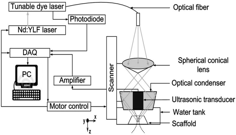
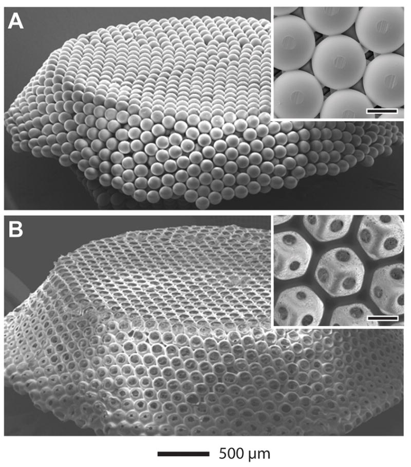
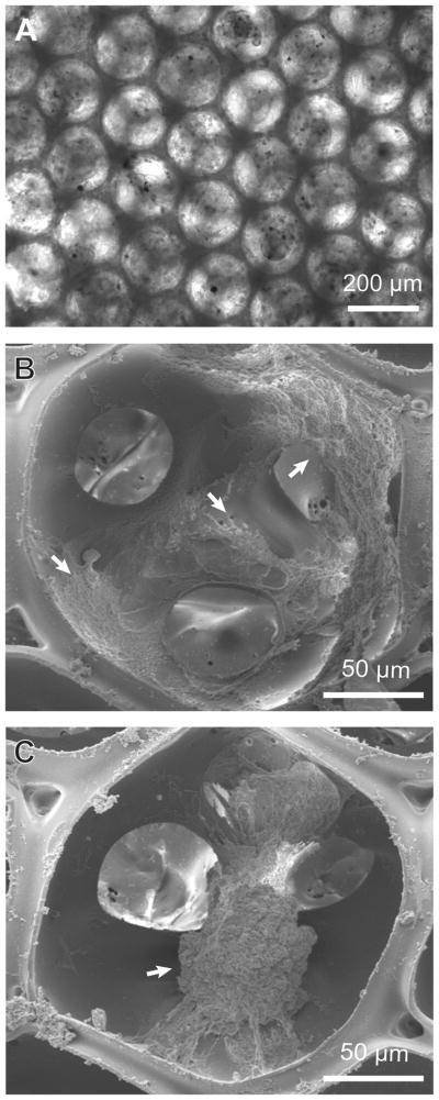
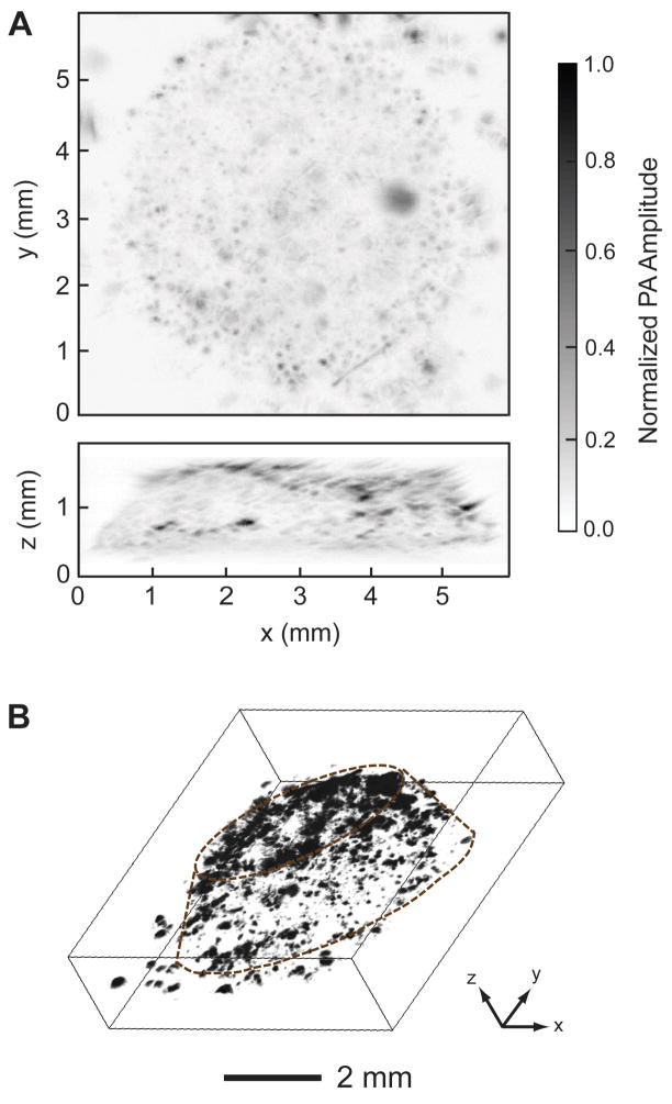
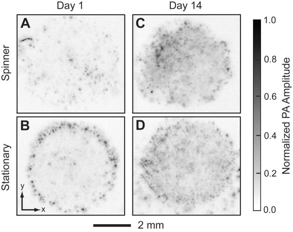
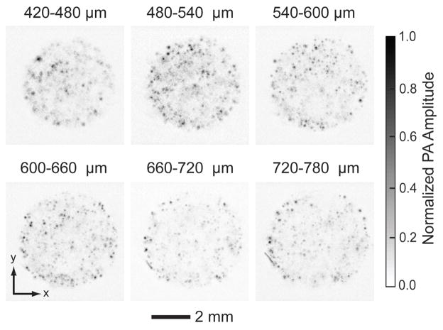
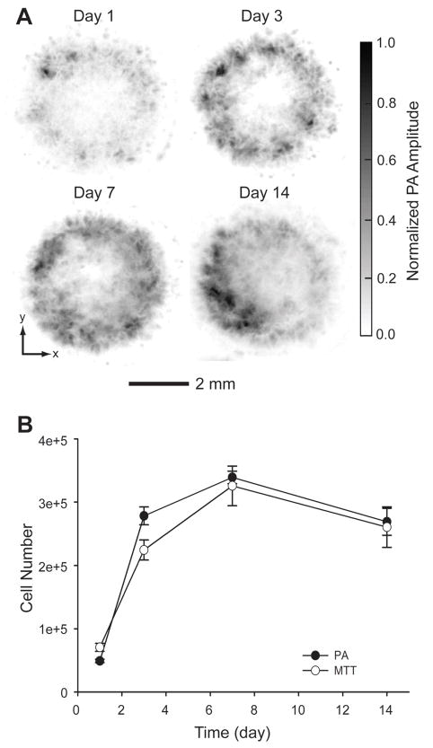
Visualizing cells in three-dimensional (3D) scaffolds has been one of the major challenges in tissue engineering. Most current imaging modalities either suffer from poor penetration depth or require exogenous contrast agents. Here, we demonstrate photoacoustic microscopy (PAM) of the spatial distribution and temporal proliferation of cells inside three-dimensional porous scaffolds with thicknesses over 1 mm. Specifically, we evaluated the effects of seeding and culture methods on the spatial distribution of melanoma cells. Spatial distribution of the cells in the scaffold was well-resolved in PAM images. Moreover, the number of cells in the scaffold was quantitatively measured from the as-obtained volumetric information. The cell proliferation profile obtained from PAM correlated well with what was obtained using the traditional 3-(4,5-dimethylthiazol-2-yl)-2,5-diphenyltetrazolium bromide (MTT) assay.
Copyright © 2010 Elsevier Ltd. All rights reserved.
Figures
Similar articles
-
Multiscale photoacoustic microscopy of single-walled carbon nanotube-incorporated tissue engineering scaffolds.Tissue Eng Part C Methods. 2012 Apr;18(4):310-7. doi: 10.1089/ten.TEC.2011.0519. Epub 2011 Dec 22. Tissue Eng Part C Methods. 2012. PMID: 22082018 Free PMC article.
-
Study on osteoblast like behavior of umbilical cord blood cells on various combinations of PLGA scaffolds prepared by salt fusion.Curr Stem Cell Res Ther. 2013 May;8(3):253-9. doi: 10.2174/1574888x11308030010. Curr Stem Cell Res Ther. 2013. PMID: 23317433
-
Functionalization of chitosan/poly(lactic acid-glycolic acid) sintered microsphere scaffolds via surface heparinization for bone tissue engineering.J Biomed Mater Res A. 2010 Jun 1;93(3):1193-208. doi: 10.1002/jbm.a.32615. J Biomed Mater Res A. 2010. PMID: 19777575
-
Tuning electrospinning parameters for production of 3D-fiber-fleeces with increased porosity for soft tissue engineering applications.Eur Cell Mater. 2011 Mar 22;21:286-303. doi: 10.22203/ecm.v021a22. Eur Cell Mater. 2011. PMID: 21432783
-
Poly(lactide-co-glycolide)/titania composite microsphere-sintered scaffolds for bone tissue engineering applications.J Biomed Mater Res B Appl Biomater. 2010 Apr;93(1):84-92. doi: 10.1002/jbm.b.31561. J Biomed Mater Res B Appl Biomater. 2010. PMID: 20091906
Cited by
-
Seeing Through the Surface: Non-invasive Characterization of Biomaterial-Tissue Interactions Using Photoacoustic Microscopy.Ann Biomed Eng. 2016 Mar;44(3):649-66. doi: 10.1007/s10439-015-1485-2. Epub 2015 Oct 15. Ann Biomed Eng. 2016. PMID: 26471785 Free PMC article. Review.
-
Non-invasive and Non-destructive Characterization of Tissue Engineered Constructs Using Ultrasound Imaging Technologies: A Review.Ann Biomed Eng. 2016 Mar;44(3):621-35. doi: 10.1007/s10439-015-1495-0. Epub 2015 Oct 30. Ann Biomed Eng. 2016. PMID: 26518412 Free PMC article. Review.
-
In vivo 3D photoacoustic and ultrasound analysis of hypopigmented skin lesions: A pilot study.Photoacoustics. 2025 Mar 8;43:100705. doi: 10.1016/j.pacs.2025.100705. eCollection 2025 Jun. Photoacoustics. 2025. PMID: 40161359 Free PMC article.
-
Fabrication of Cell Patches Using Biodegradable Scaffolds with a Hexagonal Array of Interconnected Pores (SHAIPs).Polymer (Guildf). 2014 Jan 14;55(1):445-452. doi: 10.1016/j.polymer.2013.06.019. Polymer (Guildf). 2014. PMID: 24443593 Free PMC article.
-
Photoacoustic Microscopy.Laser Photon Rev. 2013 Sep 1;7(5):10.1002/lpor.201200060. doi: 10.1002/lpor.201200060. Laser Photon Rev. 2013. PMID: 24416085 Free PMC article.
References
-
- Langer R, Vacanti JP. Tissue engineering. Science. 1993;260:920–26. - PubMed
-
- Langer R, Vacanti JP, Vacanti CA, Atala A, Freed LE, Vunjak-Novakovic G. Tissue Engineering: Biomedical Applications. Tissue Eng. 1995;1:151–61. - PubMed
-
- Vacanti JP, Langer R. Tissue engineering: the design and fabrication of living replacement devices for surgical reconstruction and transplantation. Lancet. 1999;354:S32–4. - PubMed
-
- Ma PX. Scaffolds for tissue fabrication. Mater Today. 2004;7:30–40.
-
- Hollister SJ. Porous scaffold design for tissue engineering. Nat Mater. 2005;4:518–24. - PubMed
Publication types
MeSH terms
Substances
Grants and funding
LinkOut - more resources
Full Text Sources
Medical
Molecular Biology Databases
Miscellaneous
