

Chronic label-free volumetric photoacoustic microscopy of melanoma cells in three-dimensional porous scaffolds
- PMID: 20727581
- PMCID: PMC2949455
- DOI: 10.1016/j.biomaterials.2010.07.089
Chronic label-free volumetric photoacoustic microscopy of melanoma cells in three-dimensional porous scaffolds
Abstract
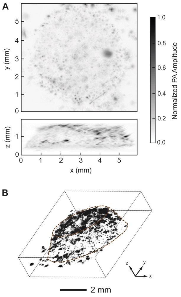
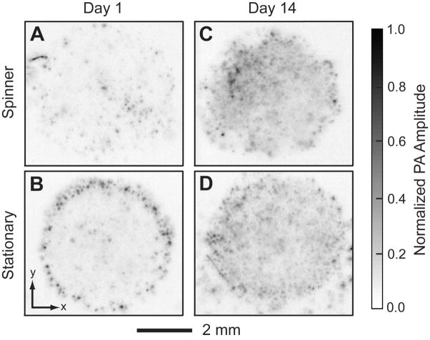
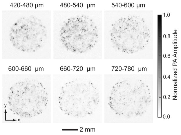
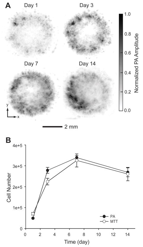
Visualizing cells in three-dimensional (3D) scaffolds has been one of the major challenges in tissue engineering. Most current imaging modalities either suffer from poor penetration depth or require exogenous contrast agents. Here, we demonstrate photoacoustic microscopy (PAM) of the spatial distribution and temporal proliferation of cells inside three-dimensional porous scaffolds with thicknesses over 1 mm. Specifically, we evaluated the effects of seeding and culture methods on the spatial distribution of melanoma cells. Spatial distribution of the cells in the scaffold was well-resolved in PAM images. Moreover, the number of cells in the scaffold was quantitatively measured from the as-obtained volumetric information. The cell proliferation profile obtained from PAM correlated well with what was obtained using the traditional 3-(4,5-dimethylthiazol-2-yl)-2,5-diphenyltetrazolium bromide (MTT) assay.
Copyright © 2010 Elsevier Ltd. All rights reserved.
Figures
References
-
- Langer R, Vacanti JP. Tissue engineering. Science. 1993;260:920–26. - PubMed
-
- Langer R, Vacanti JP, Vacanti CA, Atala A, Freed LE, Vunjak-Novakovic G. Tissue Engineering: Biomedical Applications. Tissue Eng. 1995;1:151–61. - PubMed
-
- Vacanti JP, Langer R. Tissue engineering: the design and fabrication of living replacement devices for surgical reconstruction and transplantation. Lancet. 1999;354:S32–4. - PubMed
-
- Ma PX. Scaffolds for tissue fabrication. Mater Today. 2004;7:30–40.
-
- Hollister SJ. Porous scaffold design for tissue engineering. Nat Mater. 2005;4:518–24. - PubMed
Publication types
MeSH terms
Substances
Grants and funding
LinkOut - more resources
Full Text Sources
Medical
Molecular Biology Databases
Miscellaneous
