

Warts and Yorkie mediate intestinal regeneration by influencing stem cell proliferation
- PMID: 20727758
- PMCID: PMC2955330
- DOI: 10.1016/j.cub.2010.07.041
Warts and Yorkie mediate intestinal regeneration by influencing stem cell proliferation
Abstract
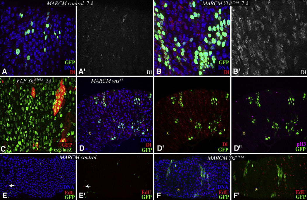
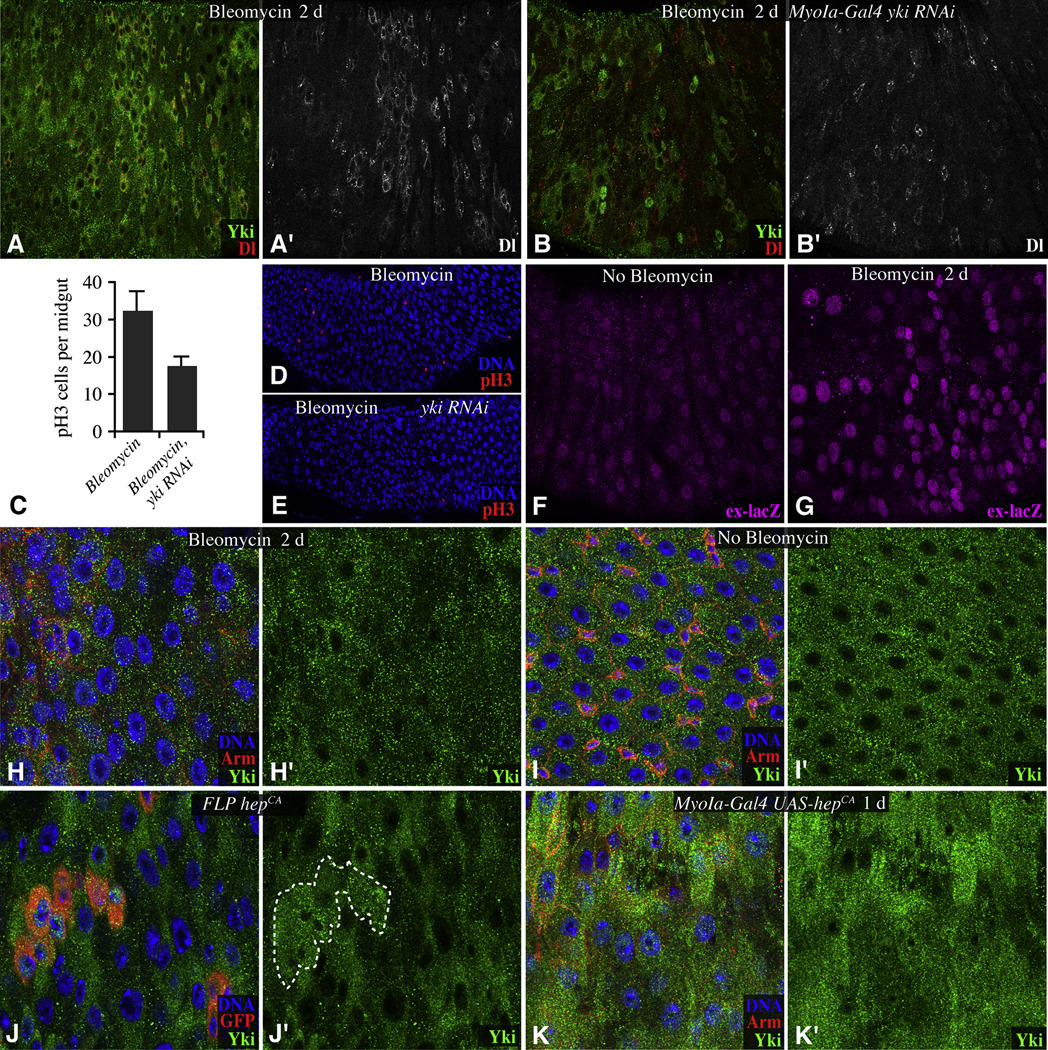
Homeostasis in the Drosophila midgut is maintained by stem cells [1, 2]. The intestinal epithelium contains two types of differentiated cells that are lost and replenished: enteroendocrine (EE) cells and enterocytes (ECs). Intestinal stem cells (ISCs) are the only cells in the adult midgut that proliferate [3, 4], and ISC divisions give rise to an ISC and an enteroblast (EB), which differentiates into an EC or an EE cell [3-5]. If the midgut epithelium is damaged, then ISC proliferation increases [6-12]. Damaged ECs express secreted ligands (Unpaired proteins) that activate Jak-Stat signaling in ISCs and EBs to promote their proliferation and differentiation [7, 9, 13, 14]. We show that the Hippo pathway components Warts and Yorkie mediate a transition from low- to high-level ISC proliferation to facilitate regeneration. The Hippo pathway regulates growth in diverse organisms and has been linked to cancer [15, 16]. Yorkie is activated in ECs in response to tissue damage or activation of the damage-sensing Jnk pathway. Activation of Yorkie promotes expression of unpaired genes and triggers a nonautonomous increase in ISC proliferation. Our observations uncover a role for Hippo pathway components in regulating stem cell proliferation and intestinal regeneration.
Copyright © 2010 Elsevier Ltd. All rights reserved.
Figures


Similar articles
-
Drosophila Pez acts in Hippo signaling to restrict intestinal stem cell proliferation.Curr Biol. 2012 Mar 6;22(5):389-96. doi: 10.1016/j.cub.2012.01.019. Epub 2012 Feb 2. Curr Biol. 2012. PMID: 22305752
-
The Hippo tumor suppressor pathway regulates intestinal stem cell regeneration.Development. 2010 Dec;137(24):4135-45. doi: 10.1242/dev.060483. Development. 2010. PMID: 21098564 Free PMC article.
-
A feedback loop between Paxillin and Yorkie sustains Drosophila intestinal homeostasis and regeneration.Nat Commun. 2025 Jan 10;16(1):570. doi: 10.1038/s41467-024-55255-1. Nat Commun. 2025. PMID: 39794306 Free PMC article.
-
Intestinal stem cell response to injury: lessons from Drosophila.Cell Mol Life Sci. 2016 Sep;73(17):3337-49. doi: 10.1007/s00018-016-2235-9. Epub 2016 May 2. Cell Mol Life Sci. 2016. PMID: 27137186 Free PMC article. Review.
-
Insect Gut Regeneration.Cold Spring Harb Perspect Biol. 2022 Feb 1;14(2):a040915. doi: 10.1101/cshperspect.a040915. Cold Spring Harb Perspect Biol. 2022. PMID: 34312250 Free PMC article. Review.
Cited by
-
Injury-induced BMP signaling negatively regulates Drosophila midgut homeostasis.J Cell Biol. 2013 Jun 10;201(6):945-61. doi: 10.1083/jcb.201302049. Epub 2013 Jun 3. J Cell Biol. 2013. PMID: 23733344 Free PMC article.
-
The Hippo pathway in intestinal regeneration and disease.Nat Rev Gastroenterol Hepatol. 2016 Jun;13(6):324-37. doi: 10.1038/nrgastro.2016.59. Epub 2016 May 5. Nat Rev Gastroenterol Hepatol. 2016. PMID: 27147489 Free PMC article. Review.
-
Hippo-Yap/Taz signaling: Complex network interactions and impact in epithelial cell behavior.Wiley Interdiscip Rev Dev Biol. 2020 May;9(3):e371. doi: 10.1002/wdev.371. Epub 2019 Dec 11. Wiley Interdiscip Rev Dev Biol. 2020. PMID: 31828974 Free PMC article. Review.
-
Brahma is essential for Drosophila intestinal stem cell proliferation and regulated by Hippo signaling.Elife. 2013 Oct 15;2:e00999. doi: 10.7554/eLife.00999. Elife. 2013. PMID: 24137538 Free PMC article.
-
Spreading the word: non-autonomous effects of apoptosis during development, regeneration and disease.Development. 2015 Oct 1;142(19):3253-62. doi: 10.1242/dev.127878. Development. 2015. PMID: 26443630 Free PMC article. Review.
References
-
- Wang P, Hou SX. Regulation of intestinal stem cells in mammals and Drosophila. J Cell Physiol. 2010;222:33–37. - PubMed
-
- Casali A, Batlle E. Intestinal stem cells in mammals and Drosophila. Cell stem cell. 2009;4:124–127. - PubMed
-
- Micchelli CA, Perrimon N. Evidence that stem cells reside in the adult Drosophila midgut epithelium. Nature. 2006;439:475–479. - PubMed
-
- Ohlstein B, Spradling A. The adult Drosophila posterior midgut is maintained by pluripotent stem cells. Nature. 2006;439:470–474. - PubMed
-
- Ohlstein B, Spradling A. Multipotent Drosophila intestinal stem cells specify daughter cell fates by differential notch signaling. Science. 2007;315:988–992. - PubMed
Publication types
MeSH terms
Substances
Grants and funding
LinkOut - more resources
Full Text Sources
Other Literature Sources
Medical
Molecular Biology Databases
Research Materials
