. 2010 Nov 1;347(1):109-21.
doi: 10.1016/j.ydbio.2010.08.014. Epub 2010 Aug 19.

Modulation of BMP signaling by Noggin is required for the maintenance of palatal epithelial integrity during palatogenesis

Affiliations

Modulation of BMP signaling by Noggin is required for the maintenance of palatal epithelial integrity during palatogenesis

Fenglei He et al. Dev Biol. .

Abstract

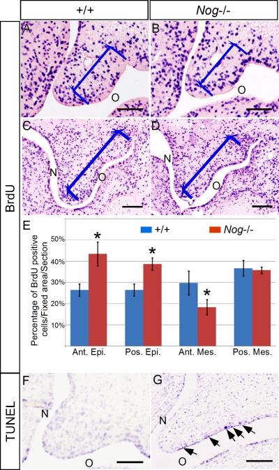
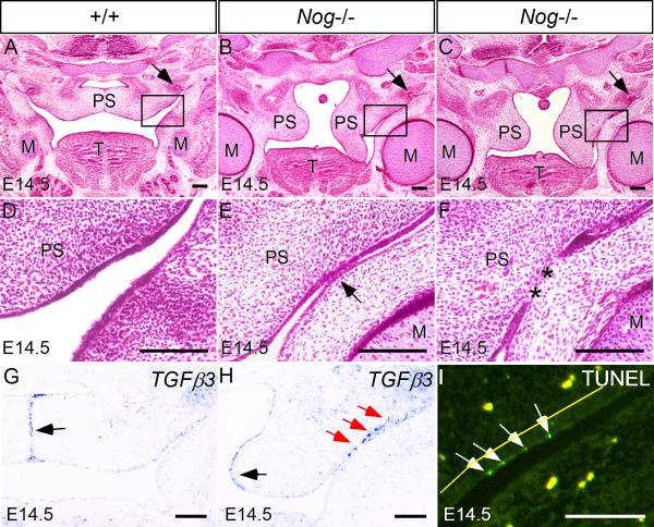
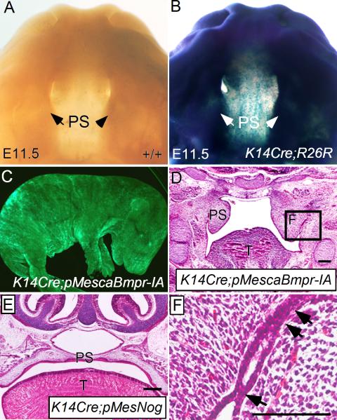
BMP signaling plays many important roles during organ development, including palatogenesis. Loss of BMP signaling leads to cleft palate formation. During development, BMP activities are finely tuned by a number of modulators at the extracellular and intracellular levels. Among the extracellular BMP antagonists is Noggin, which preferentialy binds to BMP2, BMP4 and BMP7, all of which are expressed in the developing palatal shelves. Here we use targeted Noggin mutant mice as a model for gain of BMP signaling function to investigate the role of BMP signaling in palate development. We find prominent Noggin expression in the palatal epithelium along the anterior-posterior axis during early palate development. Loss of Noggin function leads to overactive BMP signaling, particularly in the palatal epithelium. This results in disregulation of cell proliferation, excessive cell death, and changes in gene expression, leading to formation of complete palatal cleft. The excessive cell death in the epithelium disrupts the palatal epithelium integrity, which in turn leads to an abnormal palate-mandible fusion and prevents palatal shelf elevation. This phenotype is recapitulated by ectopic expression of a constitutively active form of BMPR-IA but not BMPR-IB in the epithelium of the developing palate; this suggests a role for BMPR-IA in mediating overactive BMP signaling in the absence of Noggin. Together with the evidence that overexpression of Noggin in the palatal epithelium does not cause a cleft palate defect, we conclude from our results that Noggin mediated modulation of BMP signaling is essential for palatal epithelium integrity and for normal palate development.


Figures

Figure 1
Figure 1
Expression of Noggin in the developing mouse palatal shelves. (A, B) At E11.5, Noggin mRNA is detected in the epithelium of the oral-nasal cavity, including the anterior (A) and posterior (B) palatal shelves. (C, D) In the anterior palatal shelves at E12.5, Noggin expression is detected in the nasal side and oral side palatal epithelium, but not in the MEE (C), while in the posterior palate, Noggin mRNA is restricted to the oral side palatal epithelium (D). (E-H) At E13.5, Noggin expression (arrows) is only detected in the oral side palatal epithelium (arrows) in both the anterior (E) and posterior (F) palatal shelves. LacZ reporter expression in the Nog+/- palate recapitulates the expression pattern of Noggin mRNA (G, H). (I, J) At P0, the wild type mouse develops an intact palatal shelf (I), while Nog-/- mouse exhibits a complete cleft of the secondary palate (asterisks in J) and a lack of the primary palate (arrow in J). M, Meckel's cartilage; MEE, medial edge epithelium; PP, primary palate; PS, palatal shelf; T, tongue. N designates nasal side of the palatal shelf, and O designates oral side of the palatal shelf. Scale bars represent 100 μm.
Figure 2
Figure 2
Nog-/- embryos show defective palate development. (A, C) At E13.5, the wild type palatal shelves grow vertically along the tongue in the oral-nasal cavity. (B, D) In E13.5 mutant, the anterior palatal shelves appear smaller than the wild type controls (B); in the posterior portion, the mutant palatal shelves grow shorter but slightly wider than the wild type counterparts, accompanied by ectopic cartilage formation and hyperplastic Meckel's cartilage (D). (E, G) At E14.5, the palatal shelves in wild type embryos have elevated and fused to form an intact structure above the tongue. (F, H) In the mutant at E14.5, however, the palatal shelves fail to contact each other, forming a palatal cleft (asterisk). Note that in the mutant, the anterior palatal shelves have elevated but did not make contact (F), while the posterior palatal shelves remain in a vertical position, and show abnormal fusion (arrow) with the mandible (H). M, Meckel's cartilage; MNP, medial nasal process; PS, palatal shelf; T, tongue. Scale bars represent 200 μm.
Figure 3
Figure 3
Noggin deficiency alters BMP/Smad signaling activity in the developing palate. (A, C, E, G) In the wild type control at E13.5, pSmad1/5/8 is detected in both the anterior and posterior palate. In the anterior palate, pSmad1/5/8 signal is found at high levels in the mesenchyme but is sparse in the epithelium (A, C); in the posterior palate, pSmad1/5/8/ activity is mainly restricted in the nasal side palatal mesenchyme, with a few positive signals in the oral side palatal epithelium (E, G). (B, D, F, H) In the Nog-/- palate at the comparable stage, Smad1/5/8 phosphorylation is enhanced in the oral side palatal epithelium, in both the anterior and posterior palate. As compared to the wild type control, the mutant anterior palatal mesenchyme exhibits downregulated pSmad1/5/8 signal, while the posterior palatal mesenchyme shows ectopic pSmad1/5/8 activity in the oral side. Note that the pSmad1/5/8 signal is significantly enhanced in the Nog-/- Meckel's cartilage (F). The straight white line in (E) and (F) divides the palatal mesenchyme into nasal and oral halves. The dash white lines in (C, D, G, H) demarcate the epithelial boundary. M, Meckel's cartilage; T, tongue. N designates nasal side of the palatal shelf, and O designates oral side of the palatal shelf. Scale bars represent 100 μm.
Figure 4
Figure 4
Gene expression in the wild type and Nog-/- palate. (A, B) At E13.5, Bmp4 is expressed in the anterior palatal mesenchyme (arrow) underlying MEE in the wild type palate (A); in the Nog-/- palate, comparable Bmp4 expression (arrow) is observed (B). (C, D) Comparable Msx1 expression is observed in the anterior palatal mesenchyme of E13.5 wild type (C) and Nog-/- embryo (D). (E, F) Shh expression (arrows) in the MEE of the anterior palatal shelves is not affected in Nog-/- embryo. (G, I) In E13.5 wild type controls, Bmp2 expression is detected in the anterior palatal mesenchyme and maxillary region (open arrow) (G); in the posterior palate, it is expressed in the nasal side palatal mesenchyme and the MEE region (I). (H) In the Nog-/- anterior palate, Bmp2 expression is downregulated in the palatal mesenchyme (black arrow), but is not affected in the maxillary region (open arrow). (J) In the posterior palate of the mutant, Bmp2 expression is not altered in the palatal mesenchyme and the MEE, but is ectopically activated in the oral side palatal epithelium (arrows). (K, L) Shox2 expression is significantly down-regulated in the anterior palatal mesenchyme of the mutant (L), as compared to the wild type control (K). Note comparable Shox2 expression at the ventral-lateral side of the developing tongue (red arrows). PS, palatal shelf; T, tongue. Scale bars represent 100 μm.
Figure 5
Figure 5
The Nog-/- palatal shelves exhibit defective cell proliferation and excessive apoptosis. (A-D) Coronal sections of BrdU labeled palatal shelf in the wild type and Nog-/- palate at E13.5. The blue bracket defines the regions used for cell proliferation ratio statistics. (E) Statistical data analysis shows cell proliferation rate is upregulated in the mutant palatal epithelium (both anterior and posterior). Cell proliferation rate is downregulated in the mutant anterior palatal mesenchyme, but remains unaltered in the posterior palatal mesenchyme. (F, G) At E13.5, the anterior palate of mutant exhibits excessive apoptotic cells in the oral side epithelium, but not in the nasal side (G), as compared to the wild type control (F). Ant. Epi., epithelium of anterior palate; Ant. Mes., mesenchyme of anterior palate; Pos. Epi., epithelium of posterior palate; Pos. Mes., mesenchyme of posterior palate; *, P<0.01. Scale bars represent 100 μm.
Figure 6
Figure 6
Nog-/- embryo shows abnormal palate-mandible fusion in the posterior palatal region. (A, D) At E14.5, the wild type palatal shelves have elevated to a position above the tongue and merged at the midline. (B, C, E, F) In the Nog-/- embryo at the comparable stage, the palatal shelves remain in a vertical position, showing progressive adhesion (E) and fusion (F) with the mandible. The asterisks denote a confluence of mesenchyme. The presence of the maxillary molar (arrows in A, B and C) marks the level of the sections. (G, H) Tgfβ3 is expressed in the MEE of the wild type palatal shelves (G); in the Nog-/- palate, Tgfβ3 expression is not affected in the MEE (black arrow in H), but is ectopically activated in the oral side epithelium (red arrows in H) where aberrant palate-mandible adhesion occurs. (I) TUNEL assay detects ectopic apoptotic periderm cells (arrows) in the oral side palatal epithelium prior to abnormal palate-mandible adhesion. No apoptotic cells are observed in the adjacent mandible epithelium. The yellow fluorescent dots in (I) represent autofluorescence from blood cells. The yellow line marks the basement membrane between palatal mesenchyme and the basal epithelial layer. M, Meckel's cartilage; T, tongue; PS, palatal shelf. Scale bars represent 100 μm.
Figure 7
Figure 7
Ectopic activation of BMP signaling in the palatal epithelium recapitulates the palate-mandible fusion phenotype observed in Nog-/- mice (A) An E11.5 wild type embryonic head shows negative staining of β-galactosidase. (B) An E11.5 K14Cre;R26R embryonic head shows staining of β-galactosidase in the craniofacial region, including the palatal shelves. (C) An E17.5 K14Cre;pMescaBmpr-IA embryo shows GFP expression throughout the entire embryo. (D) An E14.5 K14Cre;pMescaBmpr-IA embryo exhibits abnormal palate-mandible fusion and failure in palatal shelf elevation in the posterior portion. (E) An E16.5 K14Cre;pMesNog embryo shows a normally formed palate. (F) Higher magnification of the defined area in (D), showing an abnormal palate-mandible adhesion/fusion sites (arrows). T, tongue; PS, palatal shelf. Scale bars represent 100 μm.

Similar articles

Cited by

References

    1. Alappat SR, Zhang Z, Suzuki K, Zhang X, Liu H, Jiang R, Yamada G, Chen YP. The cellular and molecular etiology of the cleft secondary palate in Fgf10 mutant mice. Dev. Biol. 2005;277:102–113. - PubMed
    1. Anderson RM, Stottmann RW, Choi M, Klingensmith J. Endogenous bone morphogenetic protein antagonists regulate mammalian neural crest generation and survival. Dev. Dyn. 2006;235:2507–2520. - PMC - PubMed
    1. Andl T, Ahn K, Kairo A, Chu EY, Wine-Lee L, Reddy ST, Croft NJ, Cebra-Thomas JA, Metzger D, Chambon P, Lyons KM, Mishina Y, Seykora JT, Crenshaw EB, 3rd., Millar SE. Epithelial Bmpr1a regulates differentiation and proliferation in postnatal hair follicles and is essential for tooth development. Development. 2004;131:2257–2268. - PubMed
    1. Bachiller D, Klingensmith J, Kemp C, Belo JA, Anderson RM, May SR, McMahon JA, McMahon AP, Harland RM, Rossant J, De Robertis EM. The organizer factors Chordin and Noggin are required for mouse forebrain development. Nature. 2000;403:658–661. - PubMed
    1. Balemans W, Van Hul W. Extracellular regulation of BMP signaling in vertebrates: a cocktail of modulators. Dev. Biol. 2002;250:231–250. - PubMed

Publication types

MeSH terms

LinkOut - more resources