. 2010;265(2):111-9.
doi: 10.1016/j.cellimm.2010.07.011. Epub 2010 Aug 1.

Differential requirement of lipid rafts for FcγRIIA mediated effector activities

Affiliations

Differential requirement of lipid rafts for FcγRIIA mediated effector activities

Joshua A Vieth et al. Cell Immunol. 2010.

Abstract

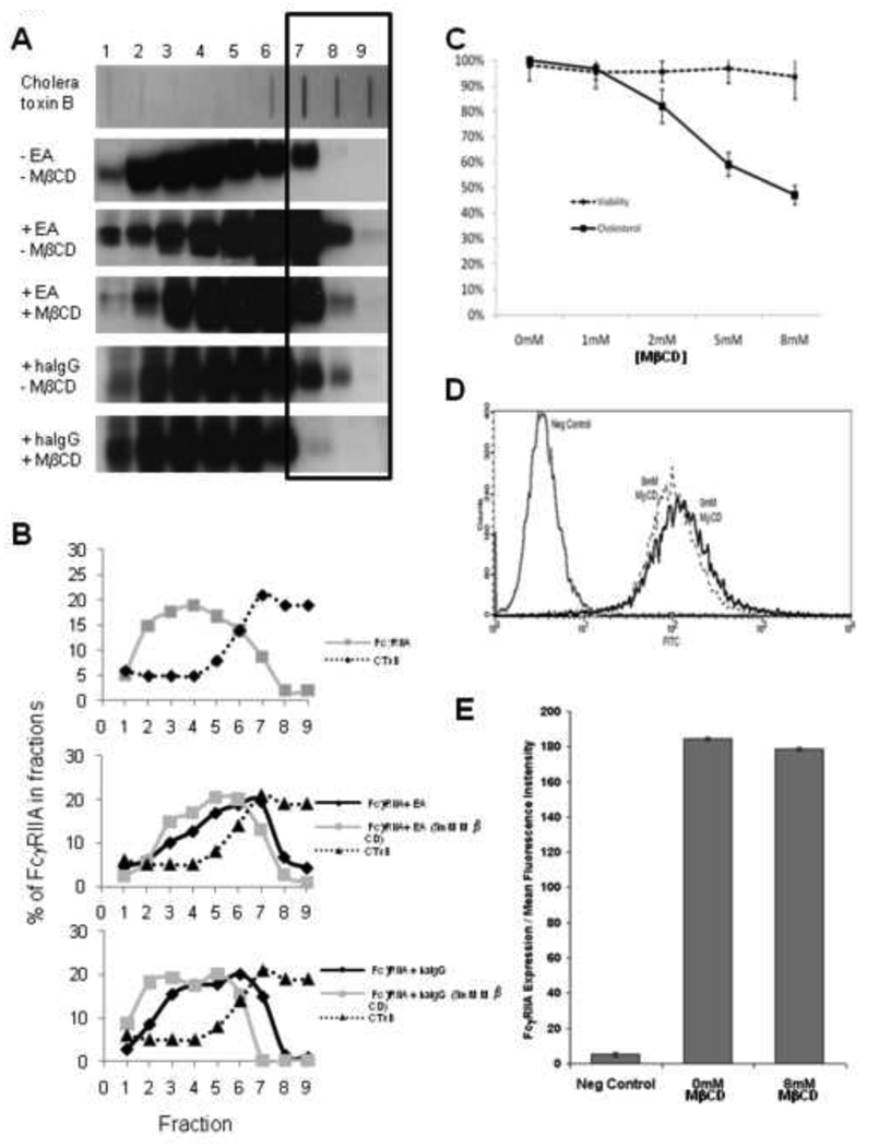
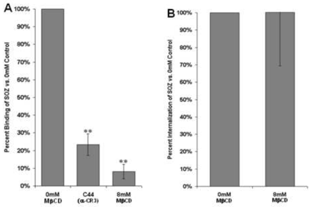
Immunoglobulin G (IgG) dependent activities are important in host defense and autoimmune diseases. Various cell types including macrophages and neutrophils contribute to pathogen destruction and tissue damage through binding of IgG to Fcγ receptors (FcγR). One member of this family, FcγRIIA, is a transmembrane glycoprotein known to mediate binding and internalization of IgG-containing targets. FcγRIIA has been observed to translocate into lipids rafts upon binding IgG-containing targets. We hypothesize that lipid rafts participate to different extents in binding and internalizing targets of different sizes. We demonstrate that disruption of lipid rafts with 8mM methyl-β-cyclodextrin (MβCD) nearly abolishes binding (91% reduction) and phagocytosis (60% reduction) of large IgG-coated targets. Conversely, binding and internalization of small IgG-complexes is less dependent on lipid rafts (49% and 17% inhibition at 8mM MβCD, respectively). These observations suggest that differences between phagocytosis and endocytosis may arise as early as the initial stages of ligand recognition.


Figures

Figure 1
Figure 1. FcγRIIA migrates into lipid rafts following activation, and this migration can be attenuated by treatment with MβCD
(A) Blot data from FcγRIIA expressing CHO cells treated with IgG coated erythrocytes (EA) (phagocytosis) or heat aggregated IgG (endocytosis). Detection of the sphingolipid GM-1 by cholera toxin B demonstrates that lipid rafts are located predominantly in fraction 6–8 (Top Row). Cross-linking with EA induces FcγRIIA to migrate into detergent insoluble fractions as shown by a shift in location toward larger numbered fractions (lanes 6–8). Treatment with MβCD inhibits the translocation. (B) The blot in A was quantitated for Cholera Toxin B binding and FcγRIIA expression using UltraQuant software. Band size and density of each lane were determined for normal cells at rest (top), the cross-linking of FcγRIIA with EA (middle), and the cross-linking of FcγRIIA with heat aggregated IgG (bottom). (C) Cell viability was assessed by Trypan blue exclusion and plasma membrane cholesterol composition via fluorescent filipin binding. As the concentration of MβCD is increased, the amount of filipin binding decreases nearly 50% while over 95% of cell remain viable. (D) Flow cytometry analysis after labeling CHO-IIA cells with anti-FcγRIIA mAB IV.3 verifies no loss of receptor expression following treatment with 8mM MβCD. (E) Data compiled from 3 experiments analyzing 10,000 cells each by flow cytometry.
Figure 2
Figure 2. Disruption of lipid rafts inhibits binding of both large IgG coated targets and small IgG complexes
(A) Representative micrographs displaying the concentration-dependent loss of binding following MβCD exposure. Small dark objects clustered around the cells are IgG-opsonized beads (OB). Blocking with IV.3 (last panel) verifies observed results are due to FcγRIIA-related activity. (B) (Top) Representative histogram demonstrating a loss in FITC-IgG signal in response to 8mM MβCD treatment, suggesting a loss in the number of FITC-conjugated IgG complexes bound to each cell. Blocking of the receptor with IV.3 completely diminished binding ability. (Bottom) Fluorescence micrographs depicting loss of fluorescence represented by less FITC-conjugated HaIgG bound to the cell surface following 8mM MβCD treatment or blocking with IV.3 mAb. (C) Binding of both IgG opsonized polystyrene beads and IgG complexes are inhibited by MβCD. Percent binding values are normalized to the 0mM control sample and represent percent of control binding after treatment. (Flow Cytometry: N=7; 10,000 cells/sample/condition. Microscopy: N=5, 300+ cells/sample/condition) (* p<0.05 compared to control, ** p<0.005 compared to control)
Figure 3
Figure 3. Disruption of lipid rafts inhibits phagocytosis of IgG opsonized beads but not endocytosis of IgG complexes
(A) CHO-IIA cells internalizing opsonized beads. Following internalization, extracellular beads were labeled with PE-conjugated goat anti-human IgG antibody to discriminate between internalized and non-internalized (right panel) beads (arrows pointing to external beads), with those fluorescing being located on the outside of the cell. (B) CHO-IIA cells internalizing FITC-conjugated HaIgG. As before, following internalization, extracellular HaIgG were labeled with PE-conjugated goat anti-human IgG antibody. Row 1 displays binding on ice with no internalization, as can be observed in the merged image, showing near complete colocalization with the secondary Ab. Row 2 exhibits changes in HaIgG location after allowing for internalization, as evidenced by an increased amount on FITC signal inside the cells, a lesser degree of PE staining, and very little colocalization in the merged image. Row 3 displays the effects of 8mM MβCD treatment prior to internalization, exhibiting a slight loss of internalization (increased PE colocalization), though some internalization is still taking place (arrows in merged image, FITC signal inside cell). (C) Representative histogram displaying measurement of IgG complex internalization. Non-labeled CHO-IIA cells exhibited no PE signal (grey line). A positive control kept on ice to inhibit internalization is included (bold black line). Samples treated with 8mM MβCD exhibited a higher level of PE labeling than untreated samples following 30 minutes of internalization at 37°C (dashed and dotted lines, respectively). This increase in PE following treatment represents more external IgG following endocytosis, thus signifying a decrease in the total amount of IgG complexes internalized. (D) Comparative graph displaying variances in lipid raft dependency for large and small targets. Percent total internalization is calculated as the loss of internalized targets normalized to control. (Flow Cytometry: N=6, 10,000 cells each sample condition. Microscopy: N=5, 300+ cells each sample condition)(* p<0.05 compared to control, ** p<0.005 compared to control)
Figure 4
Figure 4. Lipid rafts participate in binding but not phagocytosis triggered by CR3
(A) Binding of serum-opsonized zymosan particles (OZ) is decreased by lipid raft disruption, similar to what is observed for FcγRIIA (N=3, 300+ cells/sample/condition). (B) Internalization of serum opsonized zymosan particles determined by trypan blue staining is not affected by disrupting lipid raft with MβCD (N=3, 300+ cells/sample/condition). (* p<0.05 compared to control, ** p<0.005 compared to control)
Figure 5
Figure 5. Differentiated THP-1 cells display similar characteristics in lipid raft utilization as CHO cells
Dependency on lipid rafts for the binding (A) and internalization (B) of large opsonized beads and small IgG complexes in THP-1 cells mimicked results observed in our model CHO-IIA cells. Binding (Flow Cytometry: N=4, 10,000 cells each sample condition. Microscopy: N=3, 300+ cells/sample/condition). Internalization (Flow Cytometry: N=7, 10,000 cells each sample condition. Microscopy: N=6, 300+ cells/sample/condition). (* p<0.05 compared to control, ** p<0.005 compared to control)

Similar articles

Cited by

References

    1. Gavin AL, Barnes N, Dijstelbloem HM, Hogarth PM. Identification of the mouse IgG3 receptor: implications for antibody effector function at the interface between innate and adaptive immunity. J Immunol. 1998;160:20–23. - PubMed
    1. Joshi T, Butchar JP, Tridandapani S. Fcgamma receptor signaling in phagocytes. Int J Hematol. 2006;84:210–216. - PubMed
    1. Hulett MD, Hogarth PM. Molecular basis of Fc receptor function. Adv Immunol. 1994;57:1–127. - PubMed
    1. Ravetch JV, Kinet JP. Fc receptors. Annu Rev Immunol. 1991;9:457–492. - PubMed
    1. Ganesan LP, Joshi T, Fang H, Kutala VK, Roda J, Trotta R, Lehman A, Kuppusamy P, Byrd JC, Carson WE, Caligiuri MA, Tridandapani S. FcgammaR-induced production of superoxide and inflammatory cytokines is differentially regulated by SHIP through its influence on PI3K and/or Ras/Erk pathways. Blood. 2006;108:718–725. - PMC - PubMed

Publication types

LinkOut - more resources