. 2010 Sep 24;400(3):340-5.
doi: 10.1016/j.bbrc.2010.08.059. Epub 2010 Aug 20.

Isolation and characterization of the core single-stranded DNA-binding domain of purine-rich element binding protein B (Purβ)

Affiliations

Isolation and characterization of the core single-stranded DNA-binding domain of purine-rich element binding protein B (Purβ)

Amy E Rumora et al. Biochem Biophys Res Commun. .

Abstract

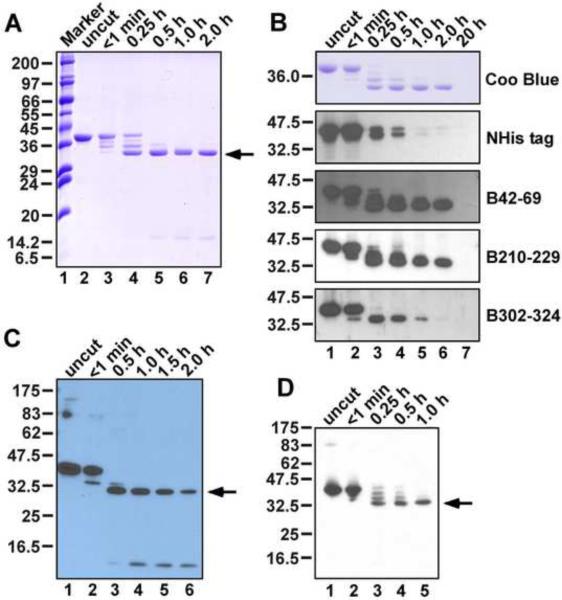
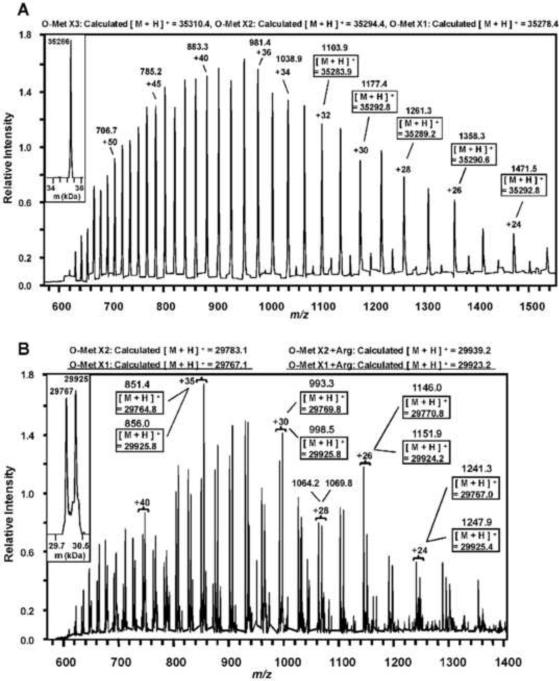
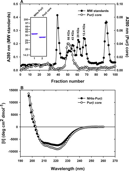
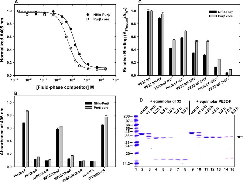
Purβ is a single-stranded nucleic acid-binding protein implicated in the injury-induced repression of genes encoding certain muscle-restricted isoforms of actin and myosin expressed in the heart, skeletal muscle, and vasculature. To better understand how the modular arrangement of the primary sequence of Purβ affects the higher order structure and function of the protein, purified recombinant Purβ was subjected to partial proteolysis in an attempt to identify a well-folded truncation protein that retained purine-rich single-stranded DNA-binding activity. Limited tryptic digestion of Purβ liberated a core ∼30kDa fragment corresponding to residues 29-305 as determined by epitope mapping and mass spectrometry. Size exclusion chromatography indicated that the isolated core fragment retains the ability to self-associate while circular dichroism analysis confirmed that the Purβ core domain is stably folded in the absence of glycine-rich N- and C-terminal sequences. Comparative DNA-binding assays revealed that the isolated core domain interacts with purine-rich cis-elements from the smooth muscle α-actin gene with similar specificity but increased affinity compared to full-length Purβ. These findings suggest that the highly conserved modular repeats of Purβ fold to form a core functional domain, which mediates the specific and high affinity binding of the protein to single-stranded DNA.


Figures

Fig. 1
Fig. 1
Identification of a core trypsin-resistant domain in Purβ. (A) Recombinant NHis-Purβ at 3.0 μM was combined with trypsin at 10:1 mass ratio. Aliquots were removed at the indicated time points. Proteolytic fragments were separated by 12% SDS-PAGE under reducing conditions and visualized by staining with Coomassie® Brilliant Blue. (B) Tryptic digestion mixtures were separated by 10% SDS-PAGE and protein fragments were either stained with Coomassie® Brilliant Blue (Coo Blue) or transferred to a PVDF membrane for immunoblotting with antibodies recognizing the NHis tag or Purβ epitopes B42–69, B210–229, or B302–324. (C) NHis-Purβ at 9.0 nM was incubated with trypsin at a 1:10 mass ratio. Aliquots were removed at the indicated time points and subjected to reducing SDS-PAGE and western blotting using a primary antibody recognizing the 210–229 epitope. (D) A tryptic digest of NHis-Purβ was subjected to southwestern blotting with a SMαA-derived probe. The arrow in panels A, C, and D identifies the core ~30 kDa fragment liberated by trypsin digestion.
Fig. 2
Fig. 2
Intact protein nano-ESI LC-MS spectra of NHis-Purβ and the core tryptic fragment. A basic deconvolution of the acquired spectra for full-length NHis-Purβ (A) and the core fragment (B) was performed using ProMass (version 25.0.1) using an input m/z range of 500–1,550 and an output m/z range of 20,000–40,000. The insets show the deconvoluted masses for the proteins based on the composite isotopic envelopes. The spectra are labeled to denote the measured and theoretical (boxed) m/z values for a range of charge states. Note for the Purβ core fragment, tryptic digestion could lead to a ragged carboxyl-terminus with or without the protein containing Arg306. This is reflected in the overlapping isotopic envelopes and deconvoluted masses exhibiting mass differences of approximately the mass of arginine (156.1 daltons). The observed and calculated masses for full-length and core Purβ with their respective methionines variably oxidized are indicated above their respective spectra.
Fig. 3
Fig. 3
Structural analysis of the Purβ core tryptic fragment via SEC and CD spectrometry. (A) A 1.5 × 98-cm column packed with Sephacryl S200 HR resin was calibrated with six different molecular weight standards (●). Resolved peaks corresponding to BSA (66 kDa), ovalbumin (43 kDa), carbonic anhydrase (29 kDa), and cytochrome c (12.4 kDa) are highlighted. In a separate run, the elution of purified Purβ core fragment (0.5 mg loaded) was monitored at 280 nm (◯). Inset, SDS-PAGE analysis of purified proteins. (B) CD scans of full-length NHis-Purβ and the core tryptic fragment were obtained in high salt buffer. The spectrum of the core fragment is consistent with loss of putative N- and C-terminal random coil-forming sequences.
Fig. 4
Fig. 4
Assessment of the ssDNA-binding affinity and specificity of the Purβ core tryptic fragment. (A) A competition assay was conducted with 0.5 nM PE32-bF in the presence of a fixed amount of solid-phase NHis-Purβ and varying amounts of fluid-phase NHis-Purβ (●) or the core fragment (◯). Data points were fit to a four parameter logistic curve using SigmaPlot 9.0 (Systat Software, Inc.). (B, C) ELISAs were performed to compare the binding of NHis-Purβ and the core fragment to StreptaWell-immobilized DNA probes corresponding to wild-type (B) or mutated (C) SMαA cis-elements and a telomeric repeat, (TTAGGG)4. (D) Limited tryptic digestion of NHis-Purβ at 5.0 μM was carried out in the presence of equimolar dT32 (lanes 2–8) or PE32-F (lanes 9–15). Lane 1 contains SigmaMarker™ proteins. Arrow shows the core fragment.

Similar articles

Cited by

References

    1. Johnson EM. The Pur protein family: clues to function from recent studies on cancer and AIDS. Anticancer Res. 2003;23:2093–2100. - PubMed
    1. Bergemann AD, Ma ZW, Johnson EM. Sequence of cDNA comprising the human pur gene and sequence-specific single-stranded-DNA-binding properties of the encoded protein. Mol. Cell. Biol. 1992;12:5673–5682. - PMC - PubMed
    1. White MK, Johnson EM, Khalili K. Multiple roles for Puralpha in cellular and viral regulation. Cell Cycle. 2009;8:1–7. - PMC - PubMed
    1. Kelm RJ, Jr., Cogan JG, Elder PK, Strauch AR, Getz MJ. Molecular interactions between single-stranded DNA-binding proteins associated with an essential MCAT element in the mouse smooth muscle α-actin promoter. J. Biol. Chem. 1999;274:14238–14245. - PubMed
    1. Carlini LE, Getz MJ, Strauch AR, Kelm RJ., Jr. Cryptic MCAT enhancer regulation in fibroblasts and smooth muscle cells. Suppression of TEF-1 mediated activation by the single-stranded DNA-binding proteins, Purα, Purβ, and MSY1. J. Biol. Chem. 2002;277:8682–8692. - PubMed

Publication types

MeSH terms

LinkOut - more resources