. 2010 Oct 28:1358:89-101.
doi: 10.1016/j.brainres.2010.08.037. Epub 2010 Aug 20.

Regional differences in estradiol effects on numbers of HSD2-containing neurons in the nucleus of the solitary tract of rats

Affiliations

Regional differences in estradiol effects on numbers of HSD2-containing neurons in the nucleus of the solitary tract of rats

Liming Fan et al. Brain Res. .

Abstract

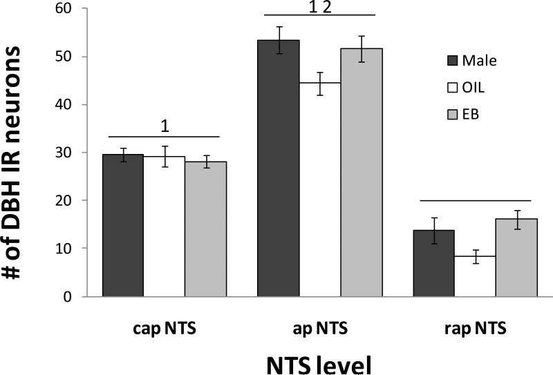
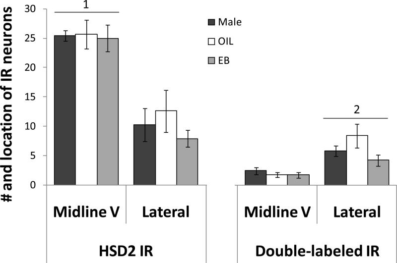
Estrogens affect body fluid balance, including sodium ingestion. Recent findings of a population of neurons in the hindbrain nucleus of the solitary tract (NTS) of rats that are activated during sodium need suggest a possible central substrate for this effect of estrogens. We used immunohistochemistry to label neurons in the NTS that express 11-β-hydroxysteroid dehydrogenase type 2 (HSD2), an enzyme that promotes aldosterone binding, in male rats, and in ovariectomized (OVX) rats given estradiol benzoate (EB) or oil vehicle (OIL). During baseline conditions, the number of HSD2 immunoreactive neurons in the NTS immediately rostral to the area postrema was greater in EB-treated OVX rats compared to those in OIL-treated OVX and male rats. A small number of HSD2 immunoreactive neurons was also labeled for dopamine-β-hydroxylase (DBH), an enzyme involved in norepinephrine biosynthesis. Double-labeled neurons in the NTS were located primarily in the more lateral portion of the HSD2 population, at the level of the area postrema in all three groups, with no sex or estrogen-mediated differences in the number of double-labeled neurons. These results suggest that two subpopulations of HSD2 neurons are present in the NTS. One subpopulation, which does not colocalize with DBH and is increased during conditions of elevated estradiol, may contribute to the effects of estrogens on sodium ingestion. The role of the other, smaller subpopulation, which colocalizes with DBH and is not affected by estradiol, remains to be determined, but one possibility is that these latter neurons are part of a larger network of catecholaminergic input to neuroendocrine neurons in the hypothalamus.


Figures

Figure 1
Figure 1
Digital photomicrographs of HSD2 and DBH immunolabeling in the NTS. Top: Examples of HSD2 immunoreactive neurons. Bottom: examples of DBH immunoreactive neurons. Scale bars - 20 μm.
Figure 2
Figure 2
Digital photomicrographs of HSD2, DBH, and double immunolabeling in the NTS. Top: a neuron labeled for HSD2 (left) and DBH (middle); double-immunolabeling (right) is indicated by yellow-orange. Bottom: neurons labeled for HSD2 (left) and DBH (middle); the field shown in the right panel includes neurons labeled only for HSD2 or DBH, as well as a neuron immunolabeled for both HSD2 and DBH. Scale bars - 20 μm.
Figure 3
Figure 3
Representative digital photomicrographs of HSD2 (top), DBH (middle), and double (bottom) immunolabeling in the capNTS. Area in box shows double immunolabeled neurons and is shown at higher magnification at the right. Green shading in the line drawing of the hindbrain indicates capNTS. dmv = dorsal motor nucleus of the vagus; scale bars - 50 μm (left panels) or 20 μm (right panels).
Figure 4
Figure 4
Representative digital photomicrographs of HSD2 (top), DBH (middle), and double (bottom) immunolabeling in the apNTS. Area in box shows double immunolabeled neurons and is shown at higher magnification at the left. Green shading in the line drawing of the hindbrain indicates apNTS. dmv = dorsal motor nucleus of the vagus; AP = area postrema; midline V = area of intensely labeled HSD2 neurons. scale bars - 50 μm (right panels) or 20 μm (left panels).
Figure 5
Figure 5
Representative digital photomicrographs of HSD2 and DBH immunolabeling in the rapNTS of a male rat (top), an OIL-treated OVX rat (middle) and an EB-treated OVX rat (bottom). Green shading in the line drawing of the hindbrain indicates rapNTS. 4V = 4th ventricle; scale bar - 50 μm.
Figure 6
Figure 6
Mean numbers of HSD2 immunolabeled neurons in the capNTS, apNTS, and rapNTS of male rats (black bars), OIL-treated OVX rats (white bars), and EB-treated OVX rats (gray bars). Overall, the number of HSD2 immunoreactive neurons in apNTS was greater than that in both capNTS and rapNTS; the number of HSD2 immunoreactive neurons in rapNTS is greater than that in capNTS (all ps<0.001). a = greater than capNTS for corresponding group (all ps<0.001); b = greater than rapNTS for corresponding group (ps<0.05, 0.001); c = greater than rapNTS in male rats (p<0.05) and in OIL-treated OVX rats (p<0.01).
Figure 7
Figure 7
Mean numbers of DBH immunolabeled neurons in the capNTS, apNTS, and rapNTS of male rats (black bars), OIL-treated OVX rats (white bars), and EB-treated OVX rats (gray bars). Overall, the number of DBH immunoreactive (IR) neurons in OIL-treated OVX rats was less than that in male rats and in EB-treated OVX rats (both ps<0.05). 1 = greater than rapNTS (both ps<0.001); 2 = greater than capNTS (p<0.001).
Figure 8
Figure 8
Mean numbers of double-immunolabeled neurons in the capNTS, apNTS, and rapNTS of male rats (black bars), OIL-treated OVX rats (white bars), and EB-treated OVX rats (gray bars). Overall, the number of double labeled neurons in apNTS was greater than that in both capNTS (p<0.01) and rapNTS (p<0.001). a = greater than rapNTS in OIL-treated OVX rats (p<0.001); b = greater than capNTS in OIL-treated OVX rats (p<0.05).
Figure 9
Figure 9
Mean numbers of HSD2 immunolabeled neurons (left) and double-labeled neurons (right) in the midline V and the more lateral portion of the apNTS of male rats (black bars), OIL-treated OVX rats (white bars), and EB-treated OVX rats (gray bars). Overall, the number of HSD2 immunoreactive neurons in the midline V was greater than that in the more lateral portion (1; p<0.001). In contrast, the number of double-labeled IR neurons in the more lateral portion of the apNTS was greater than that in the midline V (2; p<0.001).

Similar articles

Cited by

References

    1. Alves SE, Lopez V, McEwen BS, Weiland NG. Differential colocalization of estrogen receptor beta (ERbeta) with oxytocin and vasopressin in the paraventricular and supraoptic nuclei of the female rat brain: An immunocytochemical study. Proc. Natl. Acad. Sci. 1998;95:3281–3286. - PMC - PubMed
    1. Blackburn RE, Samson WK, Fulton RJ, Stricker EM, Verbalis JG. Central oxytocin and ANP receptors mediate osmotic inhibition of salt appetite in rats. Am. J. Physiol. 1995;269:R245–251. - PubMed
    1. Bossmar T, Forsling M, Akerlund M. Circulating oxytocin and vasopressin is influenced by ovarian steroid replacement in women. Acta Obstet. Gynecol. Scand. 1995;74:544–548. - PubMed
    1. Buller KM, Khanna S, Sibbald JR, Day TA. Central noradrenergic neurons signal via ATP to elicit vasopressin responses to haemorrhage. Neuroscience. 1996;73:637–642. - PubMed
    1. Chow SY, Sakai RR, Witcher JA, Adler NT, Epstein AN. Sex and sodium intake in the rat. Behav. Neurosci. 1992;106:172–180. - PubMed

Publication types