

Enhancement of virus-specific expansion of transgenic CD8 T cells in aged mice by dendritic cells
- PMID: 20728463
- PMCID: PMC2949465
- DOI: 10.1016/j.mad.2010.08.003
Enhancement of virus-specific expansion of transgenic CD8 T cells in aged mice by dendritic cells
Abstract
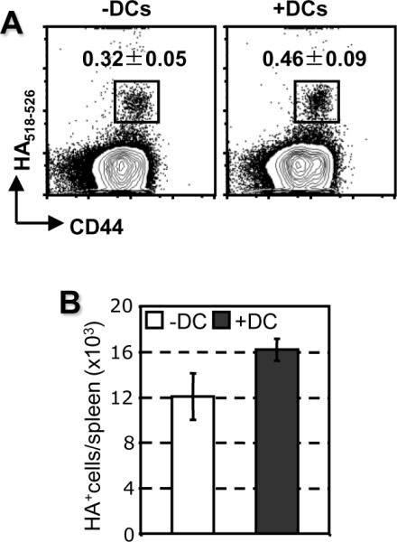
Aging is associated with a decreased CD8 T cell response to multiple antigens and to virus infection. Although both intrinsic and extrinsic factors have been shown to contribute to the decrease, the mechanisms are still largely unknown. In this study, the role of dendritic cells (DCs) in the age-associated decrease was examined. Influenza-specific TCR transgenic CD8 T cells of young mice demonstrated limited expansion in response to influenza infection when adoptively transferred to aged compared to young mice. This decreased response in aged mice could be significantly enhanced when DCs of young mice were co-transferred. Co-transfer of DCs had no impact in young recipient mice. Adoptive transfer of the DCs also increased the endogenous CD8 T cell response of intact aged mice, although to a lesser degree. These results suggest that the diminished CD8 T cell response to virus infection in aged mice is partially attributable to age-associated changes in DCs.
Copyright © 2010 Elsevier Ireland Ltd. All rights reserved.
Figures


Similar articles
-
Clearance of influenza virus from the lung depends on migratory langerin+CD11b- but not plasmacytoid dendritic cells.J Exp Med. 2008 Jul 7;205(7):1621-34. doi: 10.1084/jem.20071365. J Exp Med. 2008. PMID: 18591406 Free PMC article.
-
Limited expansion of virus-specific CD8 T cells in the aged environment.Mech Ageing Dev. 2009 Nov-Dec;130(11-12):713-21. doi: 10.1016/j.mad.2009.08.007. Mech Ageing Dev. 2009. PMID: 19744506 Free PMC article.
-
CD8 T cell responses to influenza virus infection in aged mice.Ageing Res Rev. 2011 Sep;10(4):422-7. doi: 10.1016/j.arr.2011.02.001. Epub 2011 Feb 15. Ageing Res Rev. 2011. PMID: 21315186 Free PMC article. Review.
-
Dendritic Cells Targeting Lactobacillus plantarum Strain NC8 with a Surface-Displayed Single-Chain Variable Fragment of CD11c Induce an Antigen-Specific Protective Cellular Immune Response.Infect Immun. 2020 Jan 22;88(2):e00759-19. doi: 10.1128/IAI.00759-19. Print 2020 Jan 22. Infect Immun. 2020. PMID: 31740528 Free PMC article.
-
Dendritic cells and CD28 costimulation are required to sustain virus-specific CD8+ T cell responses during the effector phase in vivo.J Immunol. 2011 Apr 15;186(8):4599-608. doi: 10.4049/jimmunol.1001972. Epub 2011 Mar 9. J Immunol. 2011. PMID: 21389258
Cited by
-
Intrinsic defects in CD8 T cells with aging contribute to impaired primary antiviral responses.Exp Gerontol. 2013 Jun;48(6):579-86. doi: 10.1016/j.exger.2013.02.027. Epub 2013 Mar 6. Exp Gerontol. 2013. PMID: 23473930 Free PMC article.
-
Cognitive and behavioral consequences of impaired immunoregulation in aging.J Neuroimmune Pharmacol. 2012 Mar;7(1):7-23. doi: 10.1007/s11481-011-9313-4. Epub 2011 Sep 20. J Neuroimmune Pharmacol. 2012. PMID: 21932047 Review.
-
Immunosenescence and Challenges of Vaccination against Influenza in the Aging Population.Aging Dis. 2012 Feb;3(1):68-90. Epub 2011 Sep 30. Aging Dis. 2012. PMID: 22500272 Free PMC article.
-
Impact of aging on antigen presentation cell function of dendritic cells.Curr Opin Immunol. 2013 Aug;25(4):535-41. doi: 10.1016/j.coi.2013.05.016. Epub 2013 Jun 24. Curr Opin Immunol. 2013. PMID: 23806201 Free PMC article. Review.
-
The aged microenvironment contributes to the age-related functional defects of CD4 T cells in mice.Aging Cell. 2012 Oct;11(5):732-40. doi: 10.1111/j.1474-9726.2012.00836.x. Epub 2012 Jun 11. Aging Cell. 2012. PMID: 22607653 Free PMC article.
References
-
- Beharka AA, Wu D, Han SN, Meydani SN. Macrophage prostaglandin production contributes to the age-associated decrease in T cell function which is reversed by the dietary antioxidant vitamin E. Mech. Ageing Dev. 1997;93:59–77. - PubMed
-
- Chiu BC, Stolberg VR, Zhang H, Chensue SW. Increased Foxp3(+) Treg cell activity reduces dendritic cell co-stimulatory molecule expression in aged mice. Mech. Ageing Dev. 2007;128:618–627. - PubMed
-
- Donnini A, Argentati K, Mancini R, Smorlesi A, Bartozzi B, Bernardini G, Provinciali M. Phenotype, antigen-presenting capacity, and migration of antigen-presenting cells in young and old age. Exp. Gerontol. 2002;37:1097–1112. - PubMed
Publication types
MeSH terms
Grants and funding
LinkOut - more resources
Full Text Sources
Medical
Research Materials
