. 2010 Nov;47(5):948-58.
doi: 10.1016/j.bone.2010.08.009. Epub 2010 Aug 20.

Identification of cells at early and late stages of polarization during odontoblast differentiation using pOBCol3.6GFP and pOBCol2.3GFP transgenic mice

Affiliations

Identification of cells at early and late stages of polarization during odontoblast differentiation using pOBCol3.6GFP and pOBCol2.3GFP transgenic mice

Anamaria Balic et al. Bone. 2010 Nov.

Abstract

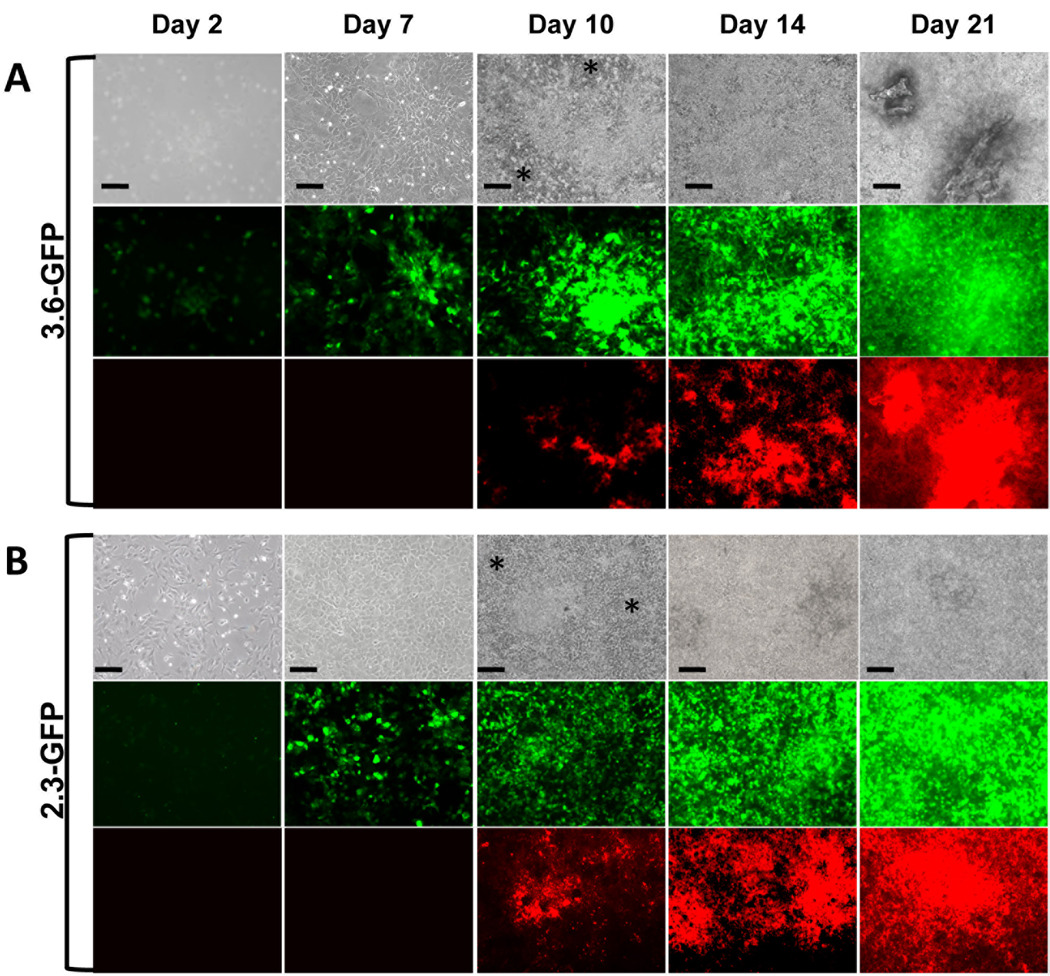
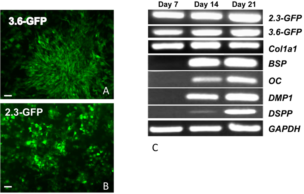
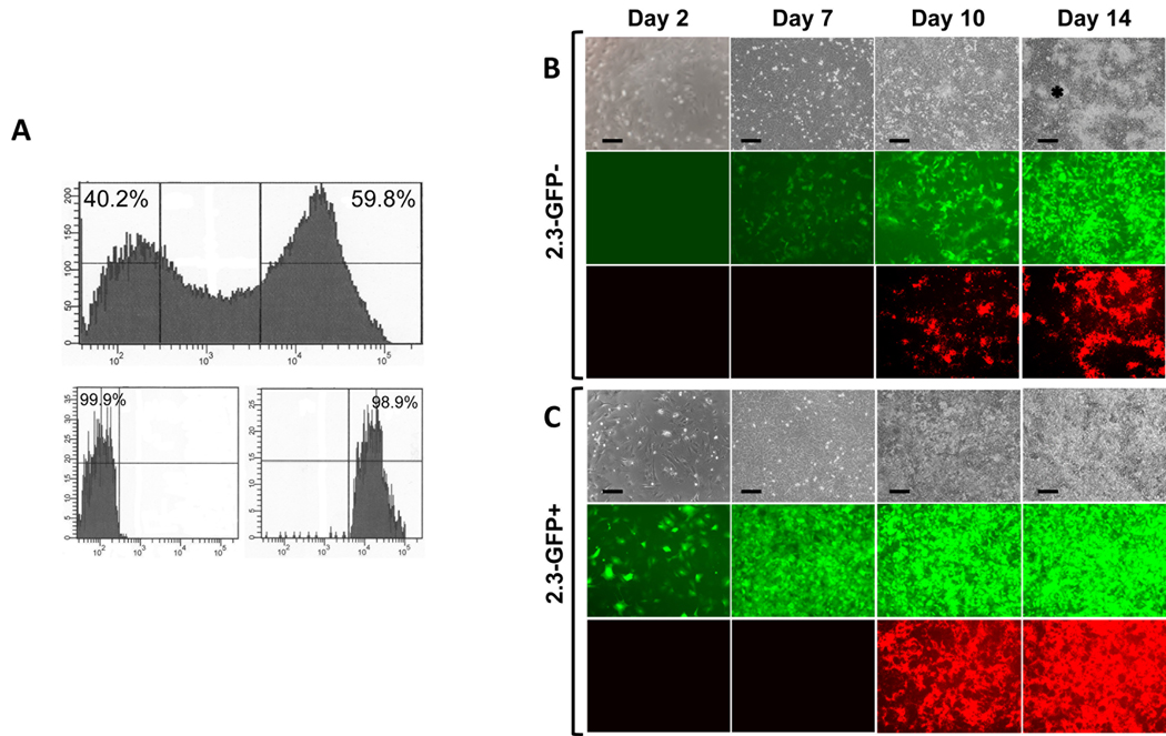
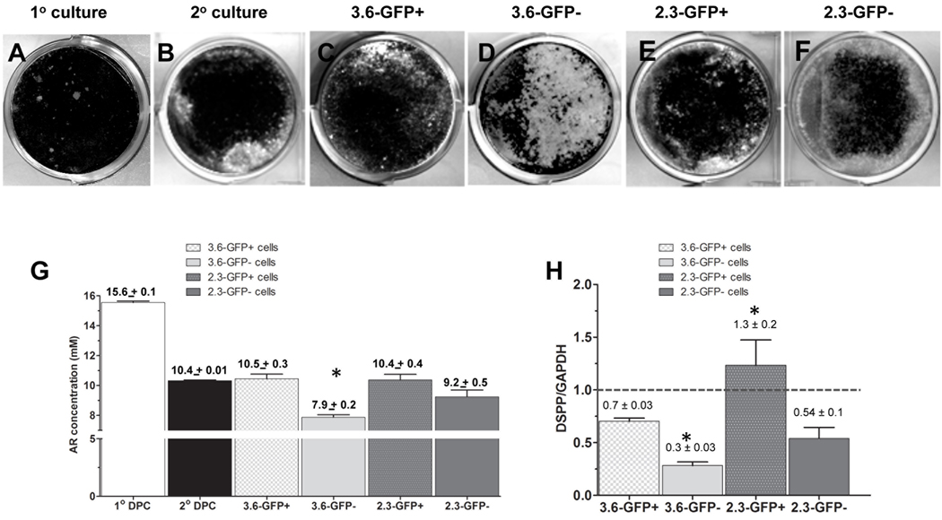
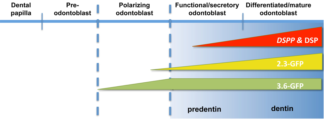
Transgenic mouse lines in which GFP expression is under the control of tissue- and stage specific promoters have provided powerful experimental tools for identification and isolation of cells at specific stage of differentiation along a lineage. In the present study, we used primary cell cultures derived from the dental pulp from pOBCol3.6GFP and pOBCol2.3GFP transgenic mice as a model to develop markers for early stages of odontoblast differentiation from progenitor cells. We analyzed the temporal and spatial expression of 2.3-GFP and 3.6-GFP during in vitro mineralization. Using FACS to separate cells based on GFP expression, we obtained relatively homogenous subpopulations of cells and analyzed their dentinogenic potentials and their progression into odontoblasts. Our observations showed that these transgenes were activated before the onset of matrix deposition and in cells at different stages of polarization. The 3.6-GFP transgene was activated in cells in early stages of polarization, whereas the 2.3-GFP transgene was activated at a later stage of polarization just before or at the time of formation of secretory odontoblast.


Figures

Figure 1
Figure 1. Expression of 3.6-GFP and 2.3-GFP transgenes in primary dental pulp cultures during in vitro mineralization and dentinogenesis
Primary dental pulp cultures obtained from pOBCol3.6GFP (A) and pOBCol2.3GFP (B) transgenic animals grown for 21 days in culture. Images of the same areas in cultures at different time points analyzed under phase contrast (upper rows in A and B), epifluorescent light using filters for GFPtpz and GFPemd for detection of GFP (middle rows in A and B) and epifluorescent light using TRITC Red filter for detection of XO staining (lower rows in A and B). Note the presence of GFP+ cells early in the culture (day 2), in cell clusters at day 7 and in differentiating and differentiated nodules between days 10–21. Note that areas of the cultures expressing GFP at day 10 extend beyond the areas of XO staining. There is a closer correlation of GFP expression and XO staining at days 14 and 21. Note the expression of low levels of 3.6-GFP and 2.3-GFP in the inter-nodular areas (indicated by asterisc). Scale bars=100µm.
Figure 2
Figure 2. Expression of early and late markers of mineralization in primary pulp cultures and their correlation with 3.6- and 2.3-GFP expression
A and B are epifluorescent images of cultures at day 7 showing the expression of 3.6-GFP in fibroblastic cells and 2.3-GFP in cuboidal cells. Scale bars=100µm. (C) RT-PCR analysis of RNA extracted from primary dental pulp pulps at different time points from pOBCol3.6GFP and pOBCol2.3GFP mice. GFP and Col1a1 expression were detected at day 7 and increases at days 14 and 21. Markers of mineralization (BSP, OC and DMP1) and dentinogenesis (DSPP) were detected at day 14 and increased at day 21.
Figure 3
Figure 3. Comparison of behavior of 3.6-GFP+ and 3.6-GFP− populations
Primary pulp cultures from pOBCol3.6GFP were grown for 7 days and processed for FACS sorting. (A) Histogram showing that FACS sorting resulted in clear separation of 3.6-GFP+ (approximately 64%) and 3.6-GFP− (approximately 35%) populations. Histogram of the FACS re-analysis shows that the purity of isolated cell populations was higher from 97%. B and C represent images of the same areas in live cultures at different time points analyzed under phase contrast (upper rows), epifluorescent light using filters for GFPtpz (middle rows in B and C) and epifluorescent light using TRITC Red filter for detection of XO staining (lower rows in B and C). Note that in 3.6-GFP+ cultures, all cells continuously expressed this transgene. XO staining was detected at day 10 and increased at day 14. In 3.6-GFP− cultures, GFP expression was not detected at day 2. At day 7, 3.6-GFP was expressed in some cells at low intensity. 3.6-GFP was expressed in the cell clusters at day 10 and increased at day 14. XO staining was detected at day 14. Cells in the inter-nodular area are indicated by asterisk. Scale bars=100µm.
Figure 4
Figure 4. Comparison of behavior of 2.3-GFP+ and 2.3-GFP− sub-populations
Primary pulp cultures derived from pOBCo12.3GFP mice were grown for 7 days and processed for FACS. (A) Histogram showing that FACS sorting resulted in clear separation of 2.3-GFP+ (approximately 60%) and 2.3-GFP− (approximately 40%) sub-populations. Histogram of FACS re-analysis on isolated cell sub-populations showed that the purity of both was higher than 98%. B and C represent images of the same areas in live cultures at different time points analyzed under phase contrast (upper rows), epifluorescent light using filters for GFPemd (middle rows in B and C) and epifluorescent light using TRITC Red filter for detection of XO staining (lower rows in B and C). Note that in cultures from 2.3-GFP+, all cells continuously expressed this transgene. XO staining was detected at day 10 and increased at day 14. In 2.3-GFP− cultures, expression of 2.3-GFP was not detected at day 2. At day 7, 2.3-GFP was expressed in some cells. Expression of 2.3-GFP was intensified at day 10 and 14. XO staining is detected at day 10 and 14. Cells in the inter-nodular area are indicated by asterisk. Scale bars=100µm.
Figure 5
Figure 5. Differentiation potentials of cultures from Col1a1-GFP+ and Col1a1-GFP−populations
A–F represent images of von Kossa staining of mineralized tissue in cultures established from unsorted cells without re-plating (primary cultures) (A), cultures from unsorted cells that were re-plated after 7 days (secondary culture) (B), 3.6-GFP+ (C), 3.6-GFP− (D), 2.3-GFP+ (E) and 2.3-GFP− (F) cells after 21 days. (G) Histogram showing the amounts of extracted Alizarin Red staining in different cultures after 21 days. Note the decrease in the mineralization in secondary pulp cultures as compared to primary pulp cultures. The amounts of extracted Alizarin Red staining were similar between secondary unsorted cells, 3.6-GFP+, 2.3-GFP+ and 2.3-GFP− populations. There was mineralization in 3.6-GFP− cultures. Values represent the concentration of the extracted Alizarin Red calculated from the mean absorbance ± S.E. for at least three independent experiments with multiple samples in each experiment (*p<0.05). (H) Histogram showing the relative levels of DSPP after 21 days in various cultures. Note the increased levels of DSPP in cultures from 2.3-GFP+ as compared to other cultures. Also note that the relative levels of DSPP were similar in 2.3-GFP− and 3.6-GFP+ cultures. The relative levels of DSPP in 3.6-GFP cultures were lower than all other cultures. The dash line represents the DSPP/GAPDH in secondary cultures that was arbitrary set to 1. Values represent the mean ± S.E. of DSPP/GAPDH for at least three independent experiments (*p<0.05).
Figure 6
Figure 6
Schematic representation of proposed stages of activation of 3.6-GFP and 2.3-GFP transgenes during odontoblast differentiation. DSPP was used as a marker of early and later stages of mineralization.

Similar articles

Cited by

References

    1. Thesleff I, Keranen S, Jernvall J. Enamel knots as signaling centers linking tooth morphogenesis and odontoblast differentiation. Adv Dent Res. 2001;15:14–18. - PubMed
    1. Arana-Chavez VE, Massa LF. Odontoblasts: the cells forming and maintaining dentine. Int J Biochem Cell Biol. 2004;36:1367–1373. - PubMed
    1. Sloan AJ, Smith AJ. Stem cells and the dental pulp: potential roles in dentine regeneration and repair. Oral Dis. 2007;13:151–157. - PubMed
    1. Lisi S, Peterkova R, Peterka M, Vonesch JL, Ruch JV, Lesot H. Tooth morphogenesis and pattern of odontoblast differentiation. Connect Tissue Res. 2003;44 Suppl 1:167–170. - PubMed
    1. Goldberg M, Smith AJ. Cells and Extracellular Matrices of Dentin and Pulp: A Biological Basis for Repair and Tissue Engineering. Crit Rev Oral Biol Med. 2004;15:13–27. - PubMed

Publication types