

Deriving Hounsfield units using grey levels in cone beam computed tomography
- PMID: 20729181
- PMCID: PMC3520236
- DOI: 10.1259/dmfr/19603304
Deriving Hounsfield units using grey levels in cone beam computed tomography
Abstract
Objectives: an in vitro study was performed to investigate the relationship between grey levels in dental cone beam CT (CBCT) and Hounsfield units (HU) in CBCT scanners.
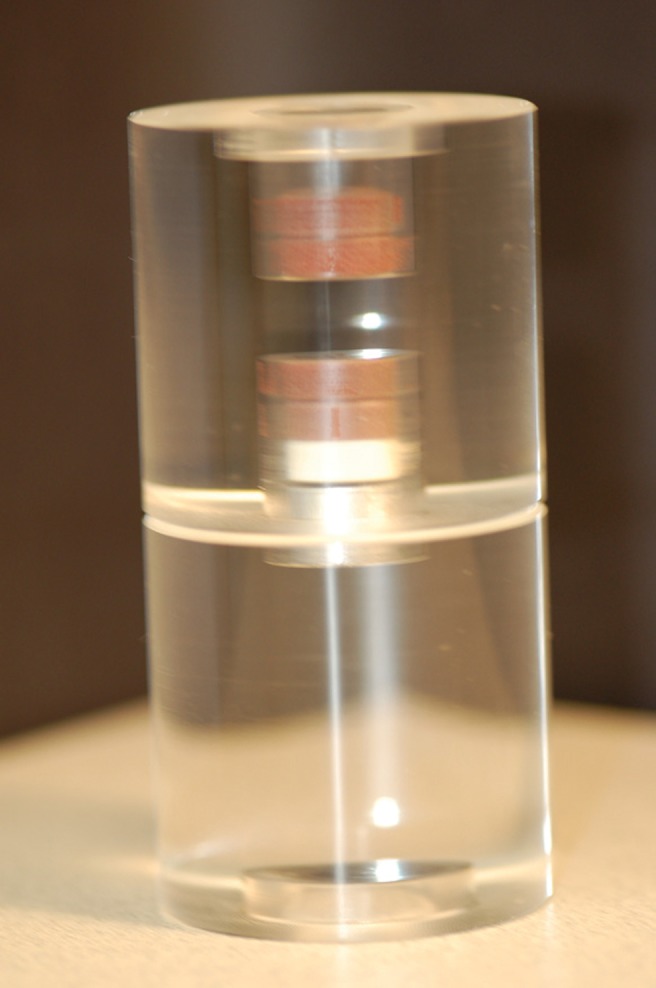
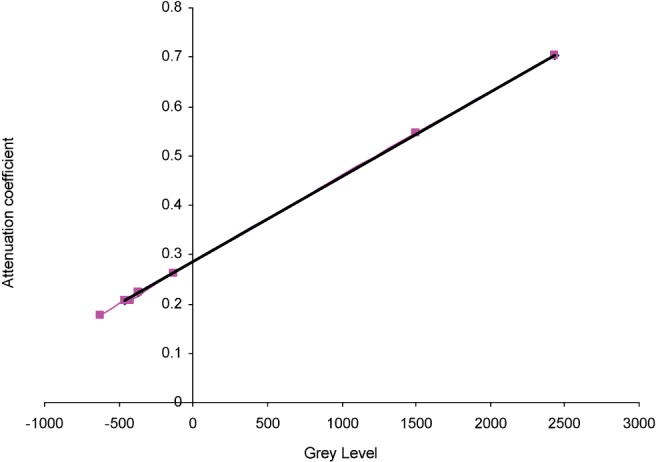
Methods: a phantom containing 8 different materials of known composition and density was imaged with 11 different dental CBCT scanners and 2 medical CT scanners. The phantom was scanned under three conditions: phantom alone and phantom in a small and large water container. The reconstructed data were exported as Digital Imaging and Communications in Medicine (DICOM) and analysed with On Demand 3D(R) by Cybermed, Seoul, Korea. The relationship between grey levels and linear attenuation coefficients was investigated.
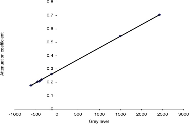
Results: it was demonstrated that a linear relationship between the grey levels and the attenuation coefficients of each of the materials exists at some "effective" energy. From the linear regression equation of the reference materials, attenuation coefficients were obtained for each of the materials and CT numbers in HU were derived using the standard equation.
Conclusions: HU can be derived from the grey levels in dental CBCT scanners using linear attenuation coefficients as an intermediate step.
Figures
Comment in
-
Deriving Hounsfield units from the grey scale of a CBCT?Dentomaxillofac Radiol. 2011 Jan;40(1):65; author reply 66. doi: 10.1259/dmfr/34858640. Dentomaxillofac Radiol. 2011. PMID: 21159918 Free PMC article. No abstract available.
Similar articles
-
Deriving Hounsfield units using grey levels in cone beam CT: a clinical application.Dentomaxillofac Radiol. 2012 Sep;41(6):500-8. doi: 10.1259/dmfr/31640433. Epub 2012 Jun 29. Dentomaxillofac Radiol. 2012. PMID: 22752324 Free PMC article.
-
Investigation of dental cone-beam CT pixel data and a modified method for conversion to Hounsfield unit (HU).Dentomaxillofac Radiol. 2018 Feb;47(2):20170321. doi: 10.1259/dmfr.20170321. Epub 2017 Nov 6. Dentomaxillofac Radiol. 2018. PMID: 29076750 Free PMC article.
-
Optimization of dental CBCT exposures through mAs reduction.Dentomaxillofac Radiol. 2015;44(9):20150108. doi: 10.1259/dmfr.20150108. Epub 2015 Jun 19. Dentomaxillofac Radiol. 2015. PMID: 26090934 Free PMC article.
-
Variability of dental cone beam CT grey values for density estimations.Br J Radiol. 2013 Jan;86(1021):20120135. doi: 10.1259/bjr.20120135. Br J Radiol. 2013. PMID: 23255537 Free PMC article.
-
Can gray values be converted to Hounsfield units? A systematic review.Dentomaxillofac Radiol. 2022 Jan 1;51(1):20210140. doi: 10.1259/dmfr.20210140. Epub 2021 Jun 19. Dentomaxillofac Radiol. 2022. PMID: 34148350 Free PMC article.
Cited by
-
Effects of photofunctionalization on early osseointegration of titanium dental implants in the maxillary posterior region: a randomized double-blinded clinical trial.Int J Implant Dent. 2021 May 10;7(1):37. doi: 10.1186/s40729-021-00318-x. Int J Implant Dent. 2021. PMID: 33969450 Free PMC article. Clinical Trial.
-
Comparative analysis of bone density measurements by using multislice spiral and cone-beam computed tomography.J Dent Sci. 2020 Sep;15(3):388-389. doi: 10.1016/j.jds.2019.09.008. Epub 2019 Dec 31. J Dent Sci. 2020. PMID: 32952898 Free PMC article. No abstract available.
-
Accuracy of computer-aided design models of the jaws produced using ultra-low MDCT doses and ASIR and MBIR.Int J Comput Assist Radiol Surg. 2018 Nov;13(11):1853-1860. doi: 10.1007/s11548-018-1809-4. Epub 2018 Jun 16. Int J Comput Assist Radiol Surg. 2018. PMID: 29909528
-
CBCT-based bone quality assessment: are Hounsfield units applicable?Dentomaxillofac Radiol. 2015;44(1):20140238. doi: 10.1259/dmfr.20140238. Dentomaxillofac Radiol. 2015. PMID: 25315442 Free PMC article. Review.
-
Proposal for a Simple and Efficient Monthly Quality Management Program Assessing the Consistency of Robotic Image-Guided Small Animal Radiation Systems.Health Phys. 2015 Nov;109(3 Suppl 3):S190-9. doi: 10.1097/HP.0000000000000323. Health Phys. 2015. PMID: 26425981 Free PMC article.
References
-
- Almog DM, Romano PR. CT-based dental imaging for implant planning and surgical guidance. NY State Dent J 2007;73:57 - PubMed
-
- Andriole KP. MDCT: a disruptive technology evolves. Imaging Economics 2004:October: www.imagingeconomics.com/
-
- Armstrong RT. Acceptability of cone beam ct vs. multi-detector CT for 3D Anatomic model construction. AAOMS 2006;64:37
-
- Mah JK, Hatcher D. Three-dimensional craniofacial imaging. Am J Orthod Dentofac Orthop 2004;126:308–309 - PubMed
-
- Quereshy FA, Savell TA, Palomo JM. Applications of cone beam computed tomography in the practice of oral and maxillofacial surgery. J Oral Maxillofac Surg 2008;66:791–796 - PubMed
Publication types
MeSH terms
LinkOut - more resources
Full Text Sources
Other Literature Sources
Medical
