

Diagnostic accuracy of cone beam computed tomography and conventional multislice spiral tomography in sheep mandibular condyle fractures
- PMID: 20729182
- PMCID: PMC3520235
- DOI: 10.1259/dmfr/29930707
Diagnostic accuracy of cone beam computed tomography and conventional multislice spiral tomography in sheep mandibular condyle fractures
Abstract
Objectives: the aim of this study was to compare diagnostic accuracy of cone beam CT (CBCT) and multislice CT in artificially created fractures of the sheep mandibular condyle.
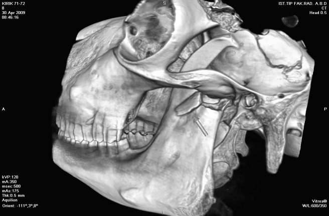
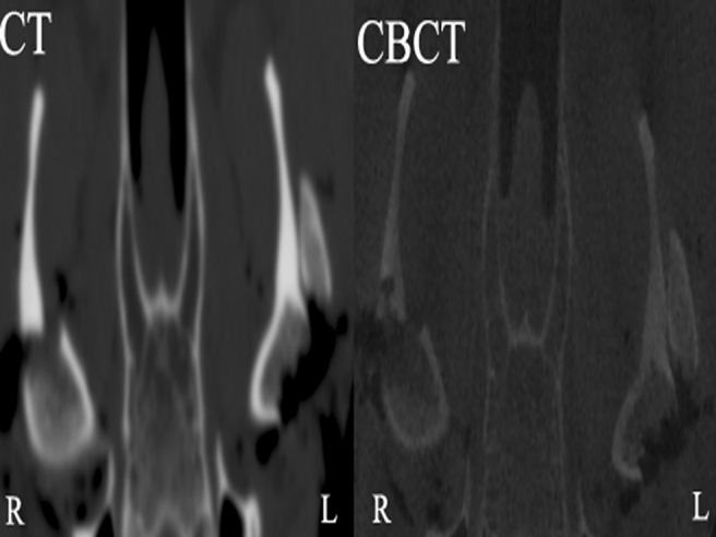
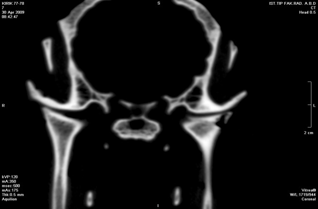
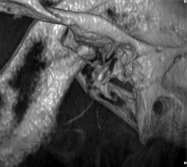
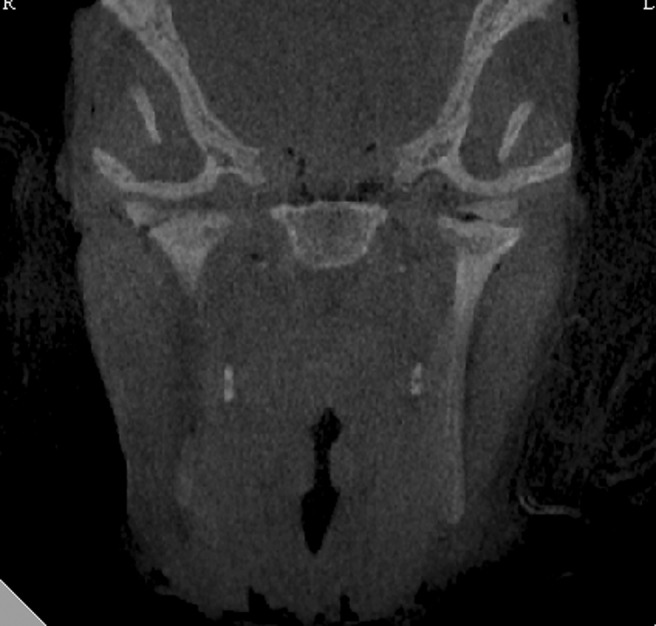
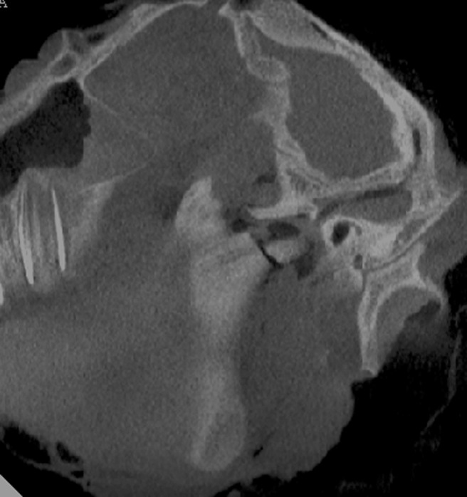
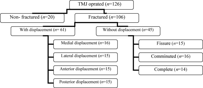
Methods: 63 full-thickness sheep heads were used in this study. Two surgeons created the fractures, which were either displaced or non-displaced. CBCT images were acquired by the NewTom 3G CBCT scanner (NIM, Verona, Italy) and CT imaging was performed using the Toshiba Aquillon multislice CT scanner (Toshiba Medical Systems, Otawara, Japan). Two-dimensional (2D) cross-sectional images and three-dimensional (3D) reconstructions were evaluated by two observers who were asked to determine the presence or absence of fracture and displacement, the type of fracture, anatomical localization and type of displacement. The naked-eye inspection during surgery served as the gold standard. Inter- and intra-observer agreements were calculated with weighted kappa statistics. The receiver operating characteristics (ROC) curve analyses were used to compare statistically the area under the curve (AUC) of both imaging modalities.
Results: kappa coefficients of intra- and interobserver agreement scores varied between 0.56 - 0.98, which were classified as moderate and excellent, respectively. There was no statistically significant difference between the imaging modalities, which were both sensitive and specific for the diagnosis of sheep condylar fractures.
Conclusions: this study confirms that CBCT is similar to CT in the diagnosis of different types of experimentally created sheep condylar fractures and can provide a cost- and dose-effective diagnostic option.
Figures
Similar articles
-
The influence of secondary reconstruction slice thickness on NewTom 3G cone beam computed tomography-based radiological interpretation of sheep mandibular condyle fractures.Oral Surg Oral Med Oral Pathol Oral Radiol Endod. 2010 Nov;110(5):638-47. doi: 10.1016/j.tripleo.2010.05.053. Oral Surg Oral Med Oral Pathol Oral Radiol Endod. 2010. PMID: 20889356
-
Interpretation of mandibular condyle fractures using 2D- and 3D-computed tomography.Braz Dent J. 2003;14(3):203-8. doi: 10.1590/s0103-64402003000300012. Epub 2004 Mar 29. Braz Dent J. 2003. PMID: 15057398
-
Detection accuracy of condylar defects in cone beam CT images scanned with different resolutions and units.Dentomaxillofac Radiol. 2014;43(3):20130414. doi: 10.1259/dmfr.20130414. Epub 2014 Jan 22. Dentomaxillofac Radiol. 2014. PMID: 24408818 Free PMC article.
-
Cone-beam computed tomography imaging of dentoalveolar and mandibular fractures.Oral Radiol. 2020 Jul;36(3):217-224. doi: 10.1007/s11282-019-00390-5. Epub 2019 May 17. Oral Radiol. 2020. PMID: 31102106 Review.
-
Reliability and accuracy of segmentation of mandibular condyles from different three-dimensional imaging modalities: a systematic review.Dentomaxillofac Radiol. 2020 Jul;49(5):20190150. doi: 10.1259/dmfr.20190150. Epub 2019 Dec 3. Dentomaxillofac Radiol. 2020. PMID: 31778321 Free PMC article.
Cited by
-
Evaluation of a low-dose protocol for cone beam computed tomography of the temporomandibular joint.Dentomaxillofac Radiol. 2020 Sep 1;49(6):20190495. doi: 10.1259/dmfr.20190495. Epub 2020 Apr 9. Dentomaxillofac Radiol. 2020. PMID: 32250642 Free PMC article.
-
High resolution MRI for quantitative assessment of inferior alveolar nerve impairment in course of mandible fractures: an imaging feasibility study.Sci Rep. 2020 Jul 14;10(1):11566. doi: 10.1038/s41598-020-68501-5. Sci Rep. 2020. PMID: 32665667 Free PMC article.
-
New evolution of cone-beam computed tomography in dentistry: Combining digital technologies.Imaging Sci Dent. 2019 Sep;49(3):179-190. doi: 10.5624/isd.2019.49.3.179. Epub 2019 Sep 24. Imaging Sci Dent. 2019. PMID: 31583200 Free PMC article. Review.
-
Clinical utility of dental cone-beam computed tomography: current perspectives.Clin Cosmet Investig Dent. 2014 Apr 2;6:29-43. doi: 10.2147/CCIDE.S41621. eCollection 2014. Clin Cosmet Investig Dent. 2014. PMID: 24729729 Free PMC article. Review.
-
Detection and classification of mandibular fractures in panoramic radiography using artificial intelligence.Dentomaxillofac Radiol. 2024 Sep 1;53(6):363-371. doi: 10.1093/dmfr/twae018. Dentomaxillofac Radiol. 2024. PMID: 38652576 Free PMC article.
References
-
- Consensus conferenceonopenorclosedmanagementofcondylarfractures 12th ICOMS. Budapest, 1995. Int J Oral Maxillofac Surg 1998;27:243–267 - PubMed
-
- Bos RR, Ward Booth RP, de Bont LG. Mandibular condyle fractures: a consensus. Br J Oral Maxillofac Surg 1999;37:87–89 - PubMed
-
- Andersson J, Hallmer F, Eriksson L. Unilateral mandibular condylar fractures: a 31-year follow-up of non-surgical treatment. Int J Oral Maxillofac Surg 2007;36:310–314 - PubMed
-
- Haug RH, Brandt MT. Closed reduction, open reduction, and endoscopic assistance: current thoughts on the management of mandibular condyle fractures. Plast Reconstr Surg 2007;120:90s–102s - PubMed
-
- Ellis E, Throckmorton GS. Treatment of mandibular condylar process fractures: biological considerations. J Oral Maxillofac Surg 2005;63:115–134 - PubMed
