

Usefulness of cone beam computed tomography in temporomandibular joints with soft tissue pathology
- PMID: 20729183
- PMCID: PMC3520237
- DOI: 10.1259/dmfr/76385066
Usefulness of cone beam computed tomography in temporomandibular joints with soft tissue pathology
Abstract
Objective: the aim of the study was to evaluate the usefulness of cone beam CT (CBCT) in temporomandibular joints (TMJs) with soft tissue pathology.
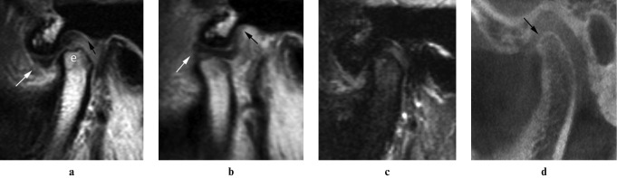
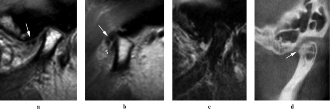
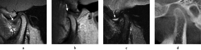
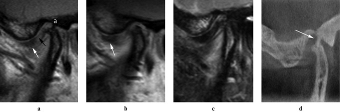
Methods: 106 TMJs of 55 patients with temporomandibular disorder (TMD) were examined by MRI and CBCT. MR images were used for the evaluation of disc displacement, disc deformity, joint effusion and obscurity of temporal posterior attachment (TPA). CBCT images were evaluated for the presence or absence of osseous abnormalities. The chi(2) test was used to analyse the association between MRI and CBCT findings.
Results: MRI of 106 TMJs revealed disc displacement, disc deformity, joint effusion and obscurity of the TPA in 68, 73, 28 and 27 joints, respectively. Of the 68 TMJs with disc displacement, anterior disc displacement without reduction (ADDWR) was seen most frequently (47/68). CBCT imaging found 65 TMJs were characterized by the presence of osseous abnormalities and were significantly associated with disc deformity and ADDWR (P < 0.05). There was no statistically significant association between the presence of joint effusion and obscurity of TPA and TMJ osseous abnormalities.
Conclusions: TMD patients with confirmed ADDWR or disc deformity on MRI are at risk of having osseous abnormalities in the TMJ and further examination with CBCT is recommended.
Figures
Similar articles
-
Relationship between temporomandibular joint space and articular disc displacement.BMC Oral Health. 2025 Apr 21;25(1):611. doi: 10.1186/s12903-025-05991-7. BMC Oral Health. 2025. PMID: 40254585 Free PMC article.
-
Inclination of the osseous components of the temporomandibular joint related with disc displacement: Magnetic resonance and cone beam computed tomography imaging-based study.Cranio. 2024 Nov;42(6):662-671. doi: 10.1080/08869634.2022.2036438. Epub 2022 Feb 14. Cranio. 2024. PMID: 35157556
-
Evaluation of the usefulness of magnetic resonance imaging in the assessment of the thickness of the roof of the glenoid fossa of the temporomandibular joint.Oral Surg Oral Med Oral Pathol Oral Radiol Endod. 2011 Oct;112(4):508-14. doi: 10.1016/j.tripleo.2011.05.013. Epub 2011 Aug 19. Oral Surg Oral Med Oral Pathol Oral Radiol Endod. 2011. PMID: 21855373
-
Ultrasonography of the temporomandibular joint: a literature review.Int J Oral Maxillofac Surg. 2009 Dec;38(12):1229-36. doi: 10.1016/j.ijom.2009.07.014. Epub 2009 Aug 22. Int J Oral Maxillofac Surg. 2009. PMID: 19700262 Review.
-
Role of magnetic resonance imaging in the clinical diagnosis of the temporomandibular joint.Cells Tissues Organs. 2005;180(1):6-21. doi: 10.1159/000086194. Cells Tissues Organs. 2005. PMID: 16088129 Review.
Cited by
-
Morphology of the Temporomandibular Joints Regarding the Presence of Osteoarthritic Changes.Int J Environ Res Public Health. 2020 Apr 23;17(8):2923. doi: 10.3390/ijerph17082923. Int J Environ Res Public Health. 2020. PMID: 32340336 Free PMC article.
-
Three-Dimensional Evaluation of Condylar Position and Joint Spaces Following Orthodontic Treatment With Quadruple Premolar Extractions.Cureus. 2024 Aug 19;16(8):e67176. doi: 10.7759/cureus.67176. eCollection 2024 Aug. Cureus. 2024. PMID: 39295726 Free PMC article.
-
Cone beam computed tomography in paediatric dentistry: overview of recent literature.Eur Arch Paediatr Dent. 2013 Jun;14(3):131-40. doi: 10.1007/s40368-013-0029-4. Eur Arch Paediatr Dent. 2013. PMID: 23564647 Review.
-
Comparison of a tridimensional cephalometric analysis performed on 3T-MRI compared with CBCT: a pilot study in adults.Prog Orthod. 2019 Oct 21;20(1):40. doi: 10.1186/s40510-019-0293-x. Prog Orthod. 2019. PMID: 31631241 Free PMC article.
-
Regional 3D superimposition to assess temporomandibular joint condylar morphology.Dentomaxillofac Radiol. 2014;43(1):20130273. doi: 10.1259/dmfr.20130273. Epub 2013 Oct 29. Dentomaxillofac Radiol. 2014. PMID: 24170802 Free PMC article.
References
-
- Okeson JP. Management of temporomandibular disorders and occlusion (6th edn). St. Louis, MO: CV Mosby Company, 2007
-
- Laskin DM, Greene CS, Hylander WL. Temporomandibular disorders: an evidence-based approach to diagnosis and treatment (1st edn). Hanover Park, IL: Quintessence Publishing, 2006
-
- Wright EF. Manual of temporomandibular disorders (2nd edn). Ames, IA: Blackwell Munksgaard, 2005
-
- Westesson PL, Katzberg RW, Tallents RH, Sanchez-Wood-worth RE, Svensson SA. CT and MR of the temporomandibular joint: comparison with autopsy specimens. AJR Am J Roentgenol 1987;148:1165–1171 - PubMed
-
- Westesson PL, Katzberg RW, Tallents RH, Sanchez-Woodworth RE, Svensson SA, Espeland MA. Temporomandibular joint: comparison of MR images with cryosectional anatomy. Radiology 1987;164:59–64 - PubMed
MeSH terms
LinkOut - more resources
Full Text Sources
Medical
