

Diagnosing common parotid tumours with magnetic resonance imaging including diffusion-weighted imaging vs fine-needle aspiration cytology: a comparative study
- PMID: 20729184
- PMCID: PMC3520240
- DOI: 10.1259/dmfr/15047967
Diagnosing common parotid tumours with magnetic resonance imaging including diffusion-weighted imaging vs fine-needle aspiration cytology: a comparative study
Abstract
Objectives: the purpose of this study was to evaluate the accuracy of MRI combined with diffusion-weighted imaging (DWI) vs fine-needle aspiration cytology (FNAC) in diagnosing common parotid masses.
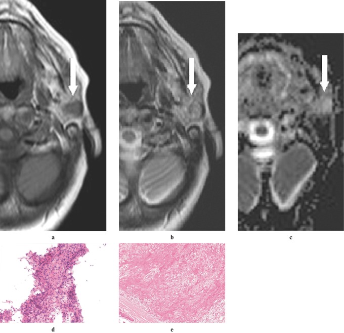
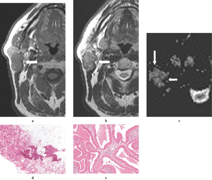
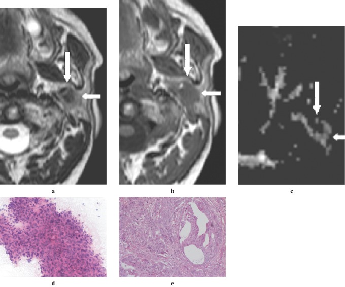
Methods: 25 consecutive patients (mean age 61 years) with parotid masses were included in this study. Informed consent and ethical approval was obtained. 22 patients underwent both MRI combined with DWI and FNAC. From DWI data, apparent diffusion coefficient maps were generated. The MRI study protocol consisted of T(1) weighted spin echo; T(2) weighted and T(2) weighted fat-suppressed turbo spin echo; DWI; and T(1) weighted fat-suppressed post-contrast images. MRI and FNAC diagnoses were compared with histopathology. Youden's index was used to compare the two methods.
Results: masses comprised eight Warthin tumours, eight adenomas (six pleomorphic adenomas, two basal cell adenomas), five carcinomas, two lipomas, one haemagioma and one benign lymphadenopathy. Technically, MRI was successful in 24 of the 25 patients (96%), FNAC was successful in 20 of the 23 patients (87.0%). The accuracy, sensitivity and specificity of MRI without DWI were 96%, 80% and 100%, respectively. Diagnostic accuracy did not increase by adding DWI to conventional MRI; however, DWI was helpful for diagnosing benign tumour histology. MRI combined with DWI was successful for determining accurate tumour typing in all benign masses except one lymphadenopathy. When FNAC had adequate material the accuracy, sensitivity and specificity were 95%, 75% and 100%, respectively. Youden's index was 0.80 for MRI and 0.75 for FNAC.
Conclusions: MRI combined with DWI seems to have similar diagnostic potential as FNAC in differentiation of benign vs malignant parotid masses.
Figures
References
-
- Wang J, Takashima S, Takayama F, Kawakami S, Saito A, Matsushita T, et al. Head and neck lesions: characterization with diffusion-weighted echo-planar MR imaging. Radiology 2001;220:621–630 - PubMed
-
- Sumi M, Takagi Y, Uetani M, Morikawa M, Hayashi K, Kabasawa H, et al. Diffusion-weighted echoplanar MR imaging of the salivary glands. AJR Am J Roentgenol 2002;178:959–965 - PubMed
-
- Tan LG, Khoo ML. Accuracy of fine needle aspiration cytology and frozen section histopathology for lesions of the major salivary glands. Ann Acad Med Singapore 2006;35:242–248 - PubMed
