. 2010 Oct 22;285(43):32906-32918.
doi: 10.1074/jbc.M110.151316. Epub 2010 Aug 20.

Further insights into the roles of GTP and the C terminus of the hepatitis C virus polymerase in the initiation of RNA synthesis

Affiliations

Further insights into the roles of GTP and the C terminus of the hepatitis C virus polymerase in the initiation of RNA synthesis

Déborah Harrus et al. J Biol Chem. .

Abstract

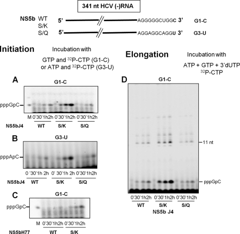
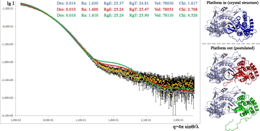
The hepatitis C virus (HCV) NS5b protein is an RNA-dependent RNA polymerase essential for replication of the viral RNA genome. In vitro and presumably in vivo, NS5b initiates RNA synthesis by a de novo mechanism. Different structural elements of NS5b have been reported to participate in RNA synthesis, especially a so-called "β-flap" and a C-terminal segment (designated "linker") that connects the catalytic core of NS5b to a transmembrane anchor. High concentrations of GTP have also been shown to stimulate de novo RNA synthesis by HCV NS5b. Here we describe a combined structural and functional analysis of genotype 1 HCV-NS5b of strains H77 (subtype 1a), for which no structure has been previously reported, and J4 (subtype 1b). Our results highlight the linker as directly involved in lifting the first boundary to processive RNA synthesis, the formation of the first dinucleotide primer. The transition from this first dinucleotide primer state to processive RNA synthesis requires removal of the linker and of the β-flap with which it is shown to strongly interact in crystal structures of HCV NS5b. We find that GTP specifically stimulates this transition irrespective of its incorporation in neosynthesized RNA.


Figures

FIGURE 1.
FIGURE 1.
Structures of H77_NS5b in complex with GTP and of J4_NS5b_S556K. NS5b is in ribbon representation and colored according to domains; fingers are in red, palm is in yellow, thumb is in blue, and linker is in light brown. A–C, shown is H77_NS5b in complex with GTP. GTP molecules are in stick representation and are colored by element; Carbon, green, nitrogen, blue; oxygen, red; phosphorus, orange. A and B, two 90° rotation views of the complex are shown. The fingertips are labeled. Orientation in A is indicated by labeling of the thumb, palm, and fingers. The linker and β-flap (see Introduction for details) are labeled in B. C, details of the H77_NS5b GTP binding site are shown. Orientation is as in B, with the palm domain removed for clarity. Residues contacting the putative priming GTP (labeled P) are shown as sticks and colored by element. All residues thus highlighted are strictly conserved among HCV strains, except the two underlined. Position 556 is always a short hydrophilic residue (Gly, Ser, Asp, or Asn), and position 446 is always an acidic residue that is conserved within genotypes (Glu for 1a, 2, and 5; Asp for 3, 4, and 6), except for subtype 1b, where 446 is a conserved Gln. Pink dashes denote hydrogen bonds. D, the same view as C is shown of the J4_NS5b_S556K structure. 2FoFc final electron density is displayed at a 1-δ level in a 1.6 Å radius around the linker and β-flap. Note that the well defined lysine 556 side chain points into the region occupied by the P GTP in C. E, shown is the primary structure of HVC_NS5b. The C-terminal transmembrane anchor, deleted from the constructs used in this study, is in dark gray.
FIGURE 2.
FIGURE 2.
Objective comparison between H77_NS5b and J4_NS5b. Shown is a ribbon view of the H77_NS5b structure in the orientation of Fig. 1A and colored after ESCET comparison with J4_NS5b using a significance level of 5 σ (see the legend to Table 3). J4_NS5b_S556K was chosen for the comparison, as it is the best defined J4_NS5b structure available (Table 4, 〈esd〉 line) and conformationally identical to other J4_NS5b structures at this significance level (Table 4, numbers in parentheses). The conformational differences between H77_NS5b and J4_NS5b indicate two major blocks in the molecule; the first (434 residues) is colored blue, and the second (89 residues) is in green, with two smaller regions (cyan and magenta); red zones indicate flexible regions. Helix A of the fingertips and helix S of the thumb are labeled.
FIGURE 3.
FIGURE 3.
Single round RNA synthesis by H77 and J4 NS5b. HCV RdRp and template RNA were preincubated for 30 min at 25 °C in the reaction mixture without NTP or with GTP or with the 2 initiating oligonucleotides. Heparin (Mr 4000–6000, 200 μg/ml) was then added followed by [α-32P]CTP and NTP needed to start the elongation. The reaction mixture was further incubated at 25 °C for 0, 5, 10, 20, and 60 min. The 32P RNA products were quantified after TCA precipitation and counted in a Wallac Counter. A, reactions were performed with G1-C and H77_NS5b (upper panel) or J4_NS5b (lower panel: empty diamonds, preincubation without NTP; filled squares, preincubation with GTP; filled triangles, preincubation with CTP and GTP). B, reactions were performed with G3-U and H77_NS5b (upper panel) or J4_NS5b (lower panel: filled squares, preincubation with GTP; filled circles, preincubation with ATP and CTP). Data were the mean of 3–6 independent experiments ± S.D.
FIGURE 4.
FIGURE 4.
Effect of high GTP concentrations on the synthesis of initiation and elongation products. A, the nucleotide sequence of the 3′ end of template RNAs G1-C, G3-U, and G3-4U is shown. B, shown is the experimental scheme. C, RNA corresponding to the 341 nt from the 3′ end of the (-) strand RNA from H77 (G1-C) or genotype 3 clone 4 (G3-U) HCV were preincubated with J4 NS5b and 0, 50, 100, 200, and 500 μm GTP for 30 min at 25 °C. [α-32P]CTP and 3′-dUTP with and without ATP were then added, and incubation continued for 2 h in the presence of 10%(v/v) DMSO. The CTP concentration was 10 μm in synthesis with G3-U and 100 μm in synthesis with G1-C. The reaction was stopped by adding 5 μl of 100 mm SDS. Five microliters were diluted in 8 μl of formamide/dyes loading buffer, and the reaction products were analyzed on 22% polyacrylamide gel. The arrowhead indicates the position of the AC dinucleotide. M is for pppGpC molecular weight marker. D, G3–4U RNA was preincubated with J4 NS5b in the same conditions as above. [α-32P]CTP with and without ATP was then added with 3′-dUTP or 3′-dGTP plus UTP, and incubation continued for 2 h in the presence of 10%(v/v) DMSO. The reaction was stopped as in C, and the synthesized RNAs were analyzed on a 20% polyacrylamide gel. M is for RNA molecular weight markers of 4, 8, and 14 nt.
FIGURE 5.
FIGURE 5.
Single-round RNA synthesis by wild type or S556 mutant NS5b. HCV RdRp and G3-U RNA were preincubated for 30 min at 25 °C in the RdRp buffer with GTP or with ATP and CTP. Heparin was then added followed by [α-32P]CTP and NTP needed to start the elongation. The reaction mixture was further incubated at 25 °C for 0, 5, 10, 20, and 60 min. The 32P RNA products were quantified after TCA precipitation and counted in a Wallac Counter. A, reactions were performed with wild type, S556K, or S556Q J4 NS5b (filled squares, preincubation with GTP; filled circles, preincubation with ATP and CTP). B, the same experiments were performed with wild type or S556K H77_NS5b. Data are the mean of six independent experiments ± S.D.
FIGURE 6.
FIGURE 6.
Effect of S556 mutations on initiation and elongation of RNA synthesis. For quantification of initiation products, wild type or mutant NS5b and 341 nt (−) RNA were incubated in the RdRp reaction buffer with 0.5 mm GTP (G1-C) or ATP (G3-U) and 10 μm [α-32P]CTP as the only added nucleotides. 10% (v/v) DMSO was also added in the experiments performed with G3-U as template (panel B). At different time points, four microliters were collected and analyzed on 22% polyacrylamide gels as described under “Experimental Procedures.” The gels were submitted to electronic autoradiography, and the radioactive bands were quantified with the Quantity one software. A, shown is J4 NS5b with G1-C. B, shown is J4 NS5b with G3-U. C, shown is H77_NS5b with G1-C. D, for quantification of elongation products, wild type or mutant J4 NS5b and G1-C were incubated in a RdRp reaction buffer with 500 μm ATP, GTP, 3′-dUTP, and 100 μm [α-32P]CTP for 2 h at 25 °C. Four microliters of the reaction were analyzed on 20% polyacrylamide gels, and the products were quantified as above.
FIGURE 7.
FIGURE 7.
Conformation of J4_NS5b in solution compared with the crystal structure and more open models. SAXS spectra measured in solution (yellow), calculated from the crystal structure J4*_O2 (blue, Platform in) or from opened models (red and green, Platform out) are shown. In a Δ55 structure (PDB code 1GX5), the thumb domain is slightly more open than in Δ21 structures (9). The red thumb model was obtained by applying this rotation to the thumb of J4*_O2 three times and moving the linker out of the catalytic core, which is the minimal deformation for the enzyme to accommodate a double-stranded RNA. The green thumb model was obtained by moving the linker further out starting from the red thumb model, which is more consistent with the elongation process occurring. The approximate expected position of the initiation platform buttressing the base of the priming nucleotide is circled in the blue thumb crystal structure and the red thumb minimally opened model.
FIGURE 8.
FIGURE 8.
Schematic of the early steps in RNA synthesis by HCV-NS5b from the HCV minus strand in vitro. The methods used in deriving this model are indicated in boxed text on the left. Increasing details are given from top to bottom, starting with the most macroscopic level of overall de novo RNA synthesis and ending with the structural elements likely involved in the salient steps of dinucleotide formation and transition to elongation. The sequence of genotype 1 (−) strand is used as an example, but the activation by GTP of the transition to elongation is independent of its incorporation in neosynthesized RNA (as shown with the G3-U and G3–4U templates under “Results”). The underlined sequence in the RNA template is paired to a complementary sequence in the conserved, stable stem-loop SL-A1 at the 3′ end of the (−) strand.

Similar articles

Cited by

References

    1. Moradpour D., Penin F., Rice C. M. (2007) Nat. Rev. Microbiol. 5, 453–463 - PubMed
    1. Gosert R., Egger D., Lohmann V., Bartenschlager R., Blum H. E., Bienz K., Moradpour D. (2003) J. Virol. 77, 5487–5492 - PMC - PubMed
    1. Ivashkina N., Wölk B., Lohmann V., Bartenschlager R., Blum H. E., Penin F., Moradpour D. (2002) J. Virol. 76, 13088–13093 - PMC - PubMed
    1. Luo G., Hamatake R. K., Mathis D. M., Racela J., Rigat K. L., Lemm J., Colonno R. J. (2000) J. Virol. 74, 851–863 - PMC - PubMed
    1. Ago H., Adachi T., Yoshida A., Yamamoto M., Habuka N., Yatsunami K., Miyano M. (1999) Structure 7, 1417–1426 - PubMed

Publication types

MeSH terms

LinkOut - more resources