. 2010 Oct 22;285(43):32824-32833.
doi: 10.1074/jbc.M110.121087. Epub 2010 Aug 19.

Inhibitors of MAPK pathway ERK1/2 or p38 prevent the IL-1{beta}-induced up-regulation of SRP72 autoantigen in Jurkat cells

Affiliations

Inhibitors of MAPK pathway ERK1/2 or p38 prevent the IL-1{beta}-induced up-regulation of SRP72 autoantigen in Jurkat cells

Victor E Arana-Argáez et al. J Biol Chem. .

Abstract

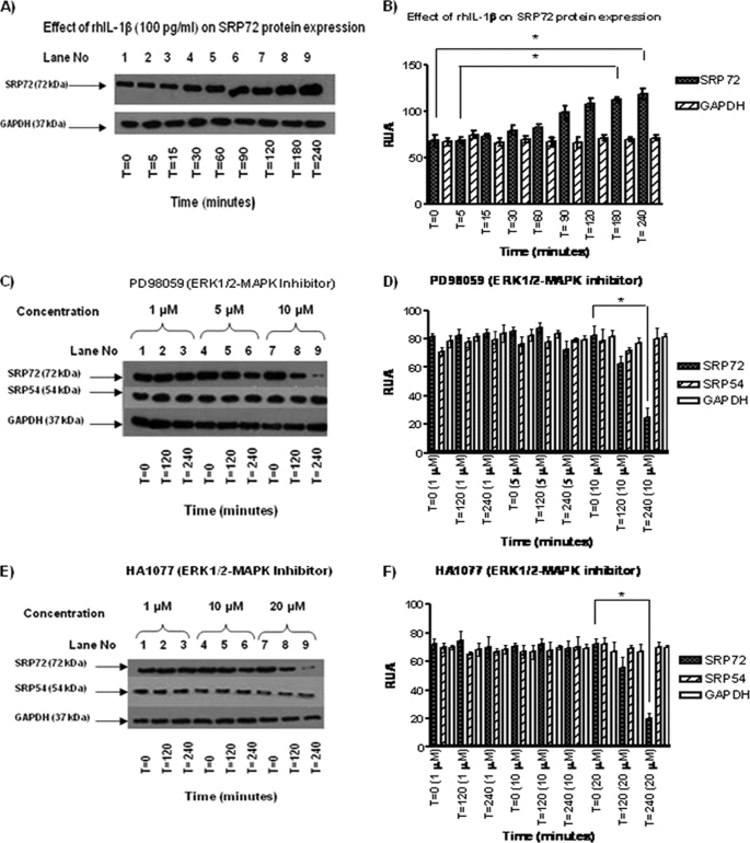
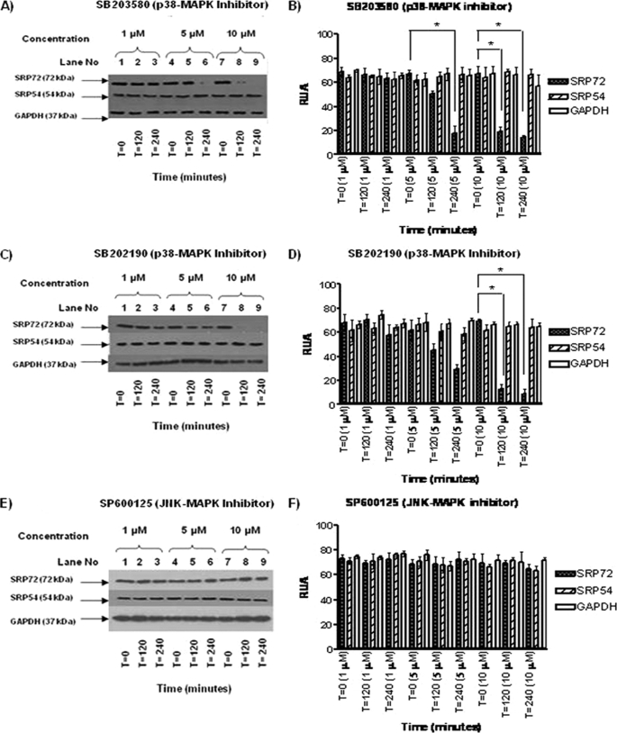
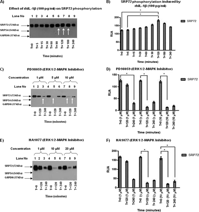
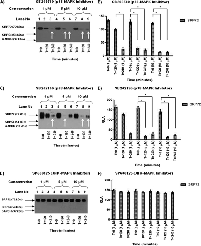
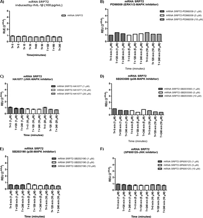
Phosphorylation is the most important post-translational event at a cellular level that is regulated by protein kinases. MAPK is a key player in the important cellular signaling pathway. It has been hypothesized that phosphorylation might have a role in the induction of break tolerance against some autoantigens such as SRP72. The aim of this study was to explore the pathways of phosphorylation and overexpression of the SRP72 polypeptide, using an in vitro model of Jurkat cells stimulated by recombinant human (rh)IL-1β in the presence of MAPK inhibitors. We used Jurkat cells as a substrate stimulated with rhIL-1β in the presence of MAPK inhibitors at different concentrations in a time course in vitro experiment by immunoprecipitation, immunoprecipitation-Western blotting, and real time PCR. Our results showed that rhIL-1β causes up-regulation of protein expression and phosphorylation of SRP72 in Jurkat cells. Inhibitors of the MAPK pathway ERK1/2 or p38α/β down-regulate the expression of SRP72 autoantigen in Jurkat cells stimulated by rhIL-1β. Our results highlight the importance of studying the pathways of activation and overexpression of autoantigens. It will be necessary to perform careful research on various kinases pathways, including MAPK in dermatomyositis and other rheumatic diseases, to help to explain the routes of activation and inhibition of autoantigens. The understanding of this process may help to develop new therapies to prevent and control the loss of tolerance toward own normal proteins.


Figures

FIGURE 1.
FIGURE 1.
A, effects of rhIL-1β on SRP72 protein expression. Jurkat cells were stimulated with rhIL-1β at different time points. WB was carried out using antibodies against human SRP72 and human GAPDH as control. We found an increase of expression of SRP72 protein at 90, 90 and 240 min (lanes 6–9), being statistically different at time 0 and 5 versus 240 and 180 min, respectively (p < 0.05). B, in contrast, GAPDH expression did not show any change. C, effect of ERK1/2 inhibitor PD98059 (1, 5, and 10 μm) on SRP72 protein expression was evaluated on Jurkat cells stimulated with rhIL-1β. The cells were harvested at 0, 120, and 240 min, and the protein expression was verified by WB using antibodies against human SRP72, human SRP54 (nonphosphorylated SRP protein), and human GAPDH as constitutive protein. A decreased SRP72 expression at 10 μm and 240 min was found. D, reduction of SRP72 expression was confirmed by RUA (closed bars) at 240 versus 0 min, 10 μm. E, effect of HA1077 (1, 10, 20 μΜ) on SRP72 protein expression by WB was evaluated at 0, 120, and 240 min. A decreased intensity of SRP72 band was found when using a concentration of 20 μm at 240 min. F, results were analyzed and RUA illustrated, finding significant results at 20 μm, 240 min (closed bars). * indicates p < 0.05. The experiments were repeated three times.
FIGURE 2.
FIGURE 2.
A, effects of p38 inhibitor SB203580 (1, 5, and 10 μm) on SRP72 protein expression was evaluated by WB using antibodies against human SRP72, SRP54, and GAPDH. A decreased intensity of SRP72 bands was noted when using the inhibitor at 5 μm concentration at 240 min (lane 6) and 10 μm at 120 and 240 min (lanes 8 and 9). B, results were analyzed and RUA illustrated, finding significant results at 5 μm, 240 versus 0 min, and 10 μm, 240 versus 0 and 120 versus 0 min, respectively, (p < 0.05). C, Jurkat cells with SB202190 at 1, 5, and 10 μm were tested, and a decreased SRP72 expression was found when using at 10 μm (lanes 8 and 9). D, results were analyzed and RUA illustrated, finding significant results at 10 μm at 240 versus 0 and 120 versus 0 min (p < 0.05). E, JNK inhibitor SP600125 (1, 5, and 10 μm, 0, 120, and 240 min); no difference in intensity of bands corresponding to SRP72, SRP54, and GAPDH was found. F, RUA of JNK inhibitor by WB was illustrated, without significant changes in protein expression of SRP72, SRP54, and GAPDH. The experiments were repeated three times.
FIGURE 3.
FIGURE 3.
A, IP of Jurkat cell lysates treated with rhIL-1β was carried out using anti-human SRP19 antibody because this is the only antibody able to immunoprecipitate the SRP complex. An anti-phosphoserine antibody was used to identify phosphorylated proteins. An increase of intensity in the SRP72 band at 90, 120, and 180 min (lanes 6–8) was found. B, results were analyzed and RUA illustrated with significant results at 120 versus 0 and versus 5 min, respectively, with p < 0.05. C, effect of ERK1/2 inhibitor PD98059 (1, 5, and 10 μm) was evaluated in SRP72-immunoprecipitated cell lysates. WB using anti-phosphoserine antibody showed a decreased SRP72 band when used as the inhibitor at concentrations of 1 μm at 240 min, 5 μm at 120 min, and 10 μm at 120 and 240 min (lanes 3, 5, 8, and 9). D, results were analyzed and RUA illustrated, finding significant results at 1 μm at 240 versus 0, 5 μm at 120 versus 0, and 10 μm at 120 versus 0 min. E, SRP72 immunoprecipitated from Jurkat cell lysate with anti-human SRP19 antibody was done to evaluate the effect of HA1077 ERK/12 inhibitor. WB using anti-phosphoserine antibody was done to identify phosphorylation status. We found a decreased intensity of SRP72 at concentration of 1 μm (120 min) and 10 and 20 μm (120 min). This finding was interesting because it suggests re-phosphorylation phenomena. F, RUA was illustrated, obtaining significant results at 5 μm at time 0 versus 120 min and 10 μm at 120 versus 0 and 240 versus 0 min (p < 0.05). * indicates p < 0.05. The experiments were repeated three times.
FIGURE 4.
FIGURE 4.
A, effect of p38 MAPK inhibitor SB203580 (1, 5, and 10 μm) on SRP72 phosphorylation was evaluated. Jurkat cells were stimulated with rhIL-1β, harvested at 0, 120, and 240 min, lysed, and immunoprecipitated using anti-human SRP19 and WB using anti-phosphoserine antibodies. A decreased intensity of SRP72 band was found when using the inhibitor at concentration of 1 μm at 240 min and 5 and 10 μm at 120 and 240 min. B, results were analyzed and RUA illustrated, finding significant results at 1 μm 240 versus 120, 5, and 10 μm at 120 versus 0 and 240 versus 0 min *, p < 0.05. C, effect of SB202190 (1, 5, and 10 μm) on SRP72 phosphorylation was tested. A decreased intensity of SRP72 expression when used at 1 μm (240 min) and 5 and 10 μm (120 and 240 min) was found. D, RUA illustrated obtaining significant results at concentration 5 and 10 μm at 120 versus 0 and 240 versus 0 min. * indicates p < 0.05. E, JNK inhibitor SP600125 (1, 5, and 10 μm) had no effect on the SRP72 intensity of bands by IP-WB using anti-phosphoserine antibody. F, results were analyzed and RUA illustrated confirming no effect of JNK inhibitors in the SRP72 expression. * indicates p < 0.05. The experiments were repeated three times.
FIGURE 5.
FIGURE 5.
We used real time RT-PCR to investigate the effects of rhIL-1β (A), PD98059 (B), HA1077 (C), SB203580 (D), SB202190 (E), and SP600125 (F) on SRP72 mRNA expression. All the samples were treated under the same experimental conditions used in protein tests done by WB, and phosphorylation was measured indirectly by IP-WB. The results did not show significant changes on the mRNA expression of SRP72 after the treatment with these inhibitors.

Similar articles

Cited by

References

    1. Bui N., Strub K. (1999) Biol. Chem. 380, 135–145 - PubMed
    1. Nagai K., Oubridge C., Kuglstatter A., Menichelli E., Isel C., Jovine L. (2003) EMBO J. 22, 3479–3485 - PMC - PubMed
    1. Buskiewicz I. A., Jöckel J., Rodnina M. V., Wintermeyer W. (2009) RNA 15, 44–54 - PMC - PubMed
    1. Fulga T. A., Sinning I., Dobberstein B., Pool M. R. (2001) EMBO J. 20, 2338–2347 - PMC - PubMed
    1. Jiang Y., Cheng Z., Mandon E. C., Gilmore R. (2008) J. Cell Biol. 180, 1149–1161 - PMC - PubMed

Publication types

MeSH terms

LinkOut - more resources