. 2010 Oct;192(20):5499-505.
doi: 10.1128/JB.00662-10. Epub 2010 Aug 20.

An extreme thermophile, Thermus thermophilus, is a polyploid bacterium

Affiliations

An extreme thermophile, Thermus thermophilus, is a polyploid bacterium

Naoto Ohtani et al. J Bacteriol. 2010 Oct.

Abstract

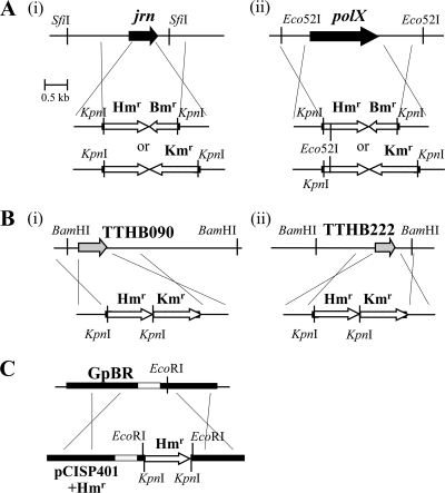
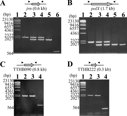
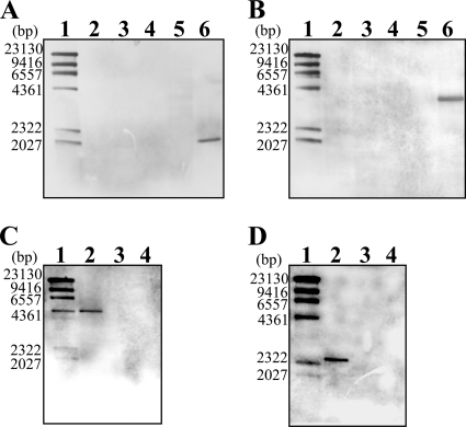
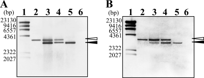
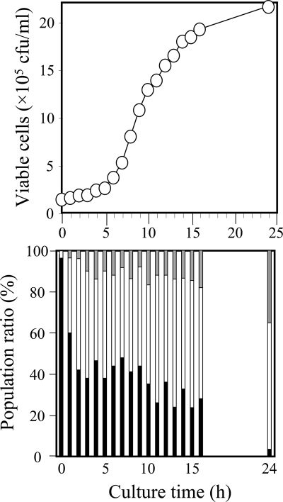
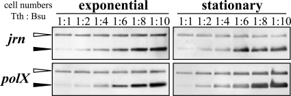
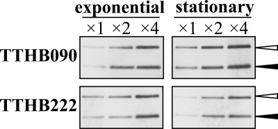
An extremely thermophilic bacterium, Thermus thermophilus HB8, is one of the model organisms for systems biology. Its genome consists of a chromosome (1.85 Mb), a megaplasmid (0.26 Mb) designated pTT27, and a plasmid (9.3 kb) designated pTT8, and the complete sequence is available. We show here that T. thermophilus is a polyploid organism, harboring multiple genomic copies in a cell. In the case of the HB8 strain, the copy number of the chromosome was estimated to be four or five, and the copy number of the pTT27 megaplasmid seemed to be equal to that of the chromosome. It has never been discussed whether T. thermophilus is haploid or polyploid. However, the finding that it is polyploid is not surprising, as Deinococcus radiodurans, an extremely radioresistant bacterium closely related to Thermus, is well known to be a polyploid organism. As is the case for D. radiodurans in the radiation environment, the polyploidy of T. thermophilus might allow for genomic DNA protection, maintenance, and repair at elevated growth temperatures. Polyploidy often complicates the recognition of an essential gene in T. thermophilus as a model organism for systems biology.


Figures

FIG. 1.
FIG. 1.
Constructs for analyses of T. thermophilus HB8 polyploidy. (A) The jrn (i) and the polX (ii) gene-null mutants in T. thermophilus HB8. The gene was deleted by replacement with the Hmr and Kmr or the Hmr and Bmr marker. The top figure represents the chromosomal region around the jrn gene, and the bottom figures represent the DNA fragments used for deletion. The crossings indicate homologous recombination. (B) The TTHB090 (i) or TTHB222 (ii) gene-null mutants to determine the quantity of the megaplasmid pTT27. In the Δjrn strain labeled by the Hmr and Bmr marker, the TTHB090 or TTHB222 gene located on the pTT27 was deleted by replacement with the Hmr and Kmr marker. The top figure represents the pTT27 region around the gene, and the bottom figure represents a DNA fragment for deletion. (C) B. subtilis possessing the Hmr sequence used for T. thermophilus HB8. This strain was used as a haploid control for the quantitation of the chromosomal copy number of T. thermophilus HB8 in Fig. 6. The top figure represents the chromosomal region around the pBR322 sequence (GpBR) of BEST7003, and the bottom figure represents the pCISP401 (pBR322 derivative) plasmid cloning Hmr. The Hmr amplified by PCR was inserted into the EcoRI site of pCISP401, and two KpnI sites were introduced just inside the resultant two EcoRI sites by PCR primers. The top and bottom shaded regions represent the tetracycline and chloramphenicol resistance genes for B. subtilis, respectively.
FIG. 2.
FIG. 2.
Confirmation of transformants by colony PCR. Colony PCR of the transformants of T. thermophilus HB8 was performed with primers located outside the deleted gene. The black triangles in the upper figures indicate the primers. The results of the jrn and the polX gene-null mutants are shown in panels A and B, respectively. Lane 1, λ/HindIII digest marker; lane 2, the transformant of the wild-type strain by the DNA fragment carrying the Hmr and Kmr marker; lanes 3 and 4, the transformants of the Hmr and Bmr strain (corresponding to the strain in lane 5) by the DNA fragment carrying the Hmr and Kmr marker; lane 5, the transformant of the wild-type strain by the DNA fragment carrying the Hmr and Bmr marker; lane 6, the wild-type strain. The results of the TTHB090 and the TTHB222 gene-null mutants are shown in panels C and D, respectively. For deletion of these genes, the Δjrn strain carrying the Hmr and Bmr marker (corresponding to the strain in lane 5 of panel A) was used as shown in Fig. 1B. Lane 1, λ/HindIII digest marker; lanes 2 and 3, the transformants of the Δjrn strain by the DNA fragment carrying the Hmr and Kmr marker; lane 4, the Δjrn strain. All of these amplified DNA fragments were checked by DNA sequencing. The numbers along the left side of the gel represent the DNA fragment size of each marker (lane 1).
FIG. 3.
FIG. 3.
Confirmation of null mutants by Southern analysis. The null mutants of T. thermophilus HB8 as shown in Fig. 2 were confirmed by Southern hybridization analysis with an inside probe for each gene. The genomic DNAs were digested by SfiI for jrn (A), Eco52I for polX (B), and BamHI for TTHB090 (C) and TTHB222 (D). (A and B) Lane 1, λ/HindIII digest marker; lane 2, the mutant carrying the Hmr and Kmr marker; lanes 3 and 4, the transformants of the Hmr- and Bmr-labeled mutant (corresponding to the strain in lane 5) by the DNA fragment carrying the Hmr and Kmr marker; lane 5, the mutant carrying the Hmr and Bmr marker; lane 6, wild type. (C and D) Lane 1, λ/HindIII digest marker; lane 2, the Δjrn strain; lanes 3 and 4, the transformant of the Δjrn strain by the DNA fragment, in which the TTHB090 or TTHB222 gene is replaced with the Hmr and Kmr marker. The numbers along the left side of the gel represent the DNA fragment size of each marker (lane 1).
FIG. 4.
FIG. 4.
Multiple chromosomes in T. thermophilus HB8. The genomic DNAs of the jrn (A)- or polX (B)-null mutants of T. thermophilus HB8 were analyzed by Southern hybridization with a probe for the Hmr marker, as described in the legend to Fig. 3. Lane 1, λ/HindIII digest marker; lane 2, the mutant carrying the Hmr and Kmr marker; lanes 3 and 4, the transformants of the Hmr- and Bmr-labeled strain (corresponding to the strain in lane 5) by the DNA fragment carrying the Hmr and Kmr marker; lane 5, the mutant carrying the Hmr and Bmr marker; lane 6, wild type. Open and closed arrowheads indicate DNA fragments containing the Hmr and Kmr and Hmr and Bmr markers, respectively. The numbers along the left side of the gel represent the DNA fragment size of each marker (lane 1). The ratios of signal intensity between DNA fragments containing the Hmr and Kmr and Hmr and Bmr markers were estimated to be approximately 1:3 for lane 3 in panel A, 2:5 for lane 4 in panel A, 5:1 for lane 3 in panel B, and 3:2 for lane 4 in panel B.
FIG. 5.
FIG. 5.
Chromosome separation kinetics. The Δjrn strain carrying both chromosomes labeled by the Hmr and Kmr or the Hmr and Bmr marker was cultured at 70°C in TR medium as described in Materials and Methods. At 0 h of culture time, the cells precultured in the presence of both Km and Bm were added to fresh antibiotic-free medium. The number of viable cells (top) was determined by counting the colonies on the antibiotic-free TR plate, and the drug resistance of each colony (bottom) was characterized by restreaking on the plate containing Km or Bm. In the bottom panel, the resistance only to Km or Bm is shown in white and gray, respectively, and that to both is shown in black.
FIG. 6.
FIG. 6.
Quantitation of chromosomal copy number of T. thermophilus HB8. The defined number of T. thermophilus HB8 Δjrn or ΔpolX cells during exponential growth and in the stationary phase was mixed with that of B. subtilis cells as a haploid control at different ratios (1:1 to 1:10), and genomic DNAs of both cells were prepared from the mixtures. The genomic DNA mixtures were digested by KpnI and used for Southern analyses with a probe for the Hmr marker. As shown in Fig. 1A and C, T. thermophilus HB8 and B. subtilis are labeled by the Hmr and Bmr and the Hmr markers, respectively. Open and closed arrowheads indicate the Hmr and Bmr and the Hmr signals, respectively.
FIG. 7.
FIG. 7.
Number of copies of the megaplasmid pTT27. Genomic DNA from the ΔTTHB090 or ΔTTHB222 strain (as described in the legend to Fig. 1B) during exponential growth or in the stationary phase was prepared, digested by KpnI, and used for Southern analyses with a probe for the Hmr marker. In the figure, ×2 and ×4 mean that 2- and 4-fold amounts of the digested DNAs were applied for electrophoresis. The strain possesses the Hmr and Bmr and the Hmr and Kmr markers replacing the jrn gene on the chromosome and the TTHB090 gene on the pTT27, respectively. Open and closed arrowheads indicate the Hmr and Kmr signal on pTT27 and the Hmr and Bmr signal on the chromosome, respectively.

Similar articles

Cited by

References

    1. Angert, E. R., and K. D. Clements. 2004. Initiation of intracellular offspring in Epulopiscium. Mol. Microbiol. 51:827-835. - PubMed
    1. Binder, B. J., and S. W. Chisholm. 1990. Relationship between DNA cycle and growth rate in Synechococcus sp. strain PCC 6301. J. Bacteriol. 172:2313-2319. - PMC - PubMed
    1. Bremer, H., and P. P. Dennis. 1996. Modulation of chemical composition and other parameters of the cell growth rate, p. 1553-1566. In F. C. Neidhardt (ed.), Escherichia coli and Salmonella. ASM Press, Washington, DC.
    1. Bresler, V., W. L. Montgomery, L. Fishelson, and P. E. Pollak. 1998. Gigantism in a bacterium, Epulopiscium fishelsoni, correlates with complex patterns in arrangement, quantity, and segregation of DNA. J. Bacteriol. 180:5601-5611. - PMC - PubMed
    1. Brouns, S. J., H. Wu, J. Akerboom, A. P. Turnbull, W. M. de Vos, and J. van der Oost. 2005. Engineering a selectable marker for hyperthermophiles. J. Biol. Chem. 280:11422-11431. - PubMed

Publication types