. 2010 Oct;192(20):5413-23.
doi: 10.1128/JB.00592-10. Epub 2010 Aug 20.

Characterization of the RNA degradosome of Pseudoalteromonas haloplanktis: conservation of the RNase E-RhlB interaction in the gammaproteobacteria

Affiliations

Characterization of the RNA degradosome of Pseudoalteromonas haloplanktis: conservation of the RNase E-RhlB interaction in the gammaproteobacteria

Soraya Aït-Bara et al. J Bacteriol. 2010 Oct.

Abstract

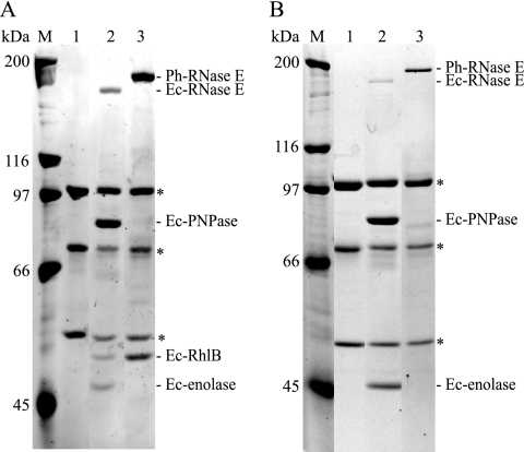
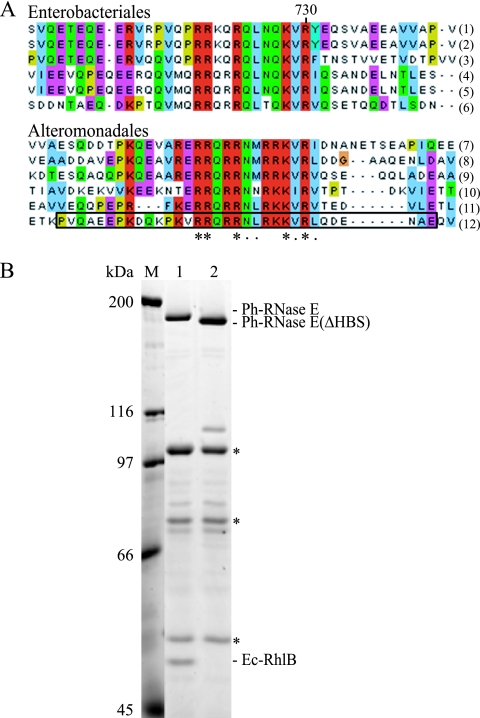
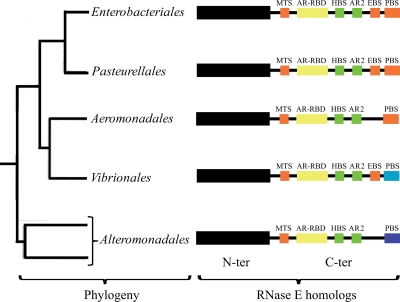
The degradosome is a multienzyme complex involved in mRNA degradation in Escherichia coli. The essential endoribonuclease RNase E contains a large noncatalytic region necessary for protein-protein interactions with other components of the RNA degradosome. Interacting proteins include the DEAD-box RNA helicase RhlB, the glycolytic enzyme enolase, and the exoribonuclease PNPase. Pseudoalteromonas haloplanktis, a psychrotolerant gammaproteobacterium distantly related to E. coli, encodes homologs of each component of the RNA degradosome. In P. haloplanktis, RNase E associates with RhlB and PNPase but not enolase. Plasmids expressing P. haloplanktis RNase E (Ph-RNase E) can complement E. coli strains lacking E. coli RNase E (Ec-RNase E). Ph-RNase E, however, does not confer a growth advantage to E. coli at low temperature. Ph-RNase E has a heterologous protein-protein interaction with Ec-RhlB but not with Ec-enolase or Ec-PNPase. The Ph-RNase E binding sites for RhlB and PNPase were mapped by deletion analysis. The PNPase binding site is located at the C-terminal end of Ph-RNase E at the same position as that in Ec-RNase E, but the sequence of the site is not conserved. The sequence of the RhlB binding site in Ph-RNase E is related to the sequence in Ec-RNase E. Together with the heterologous interaction between Ph-RNase E and Ec-RhlB, our results suggest that the underlying structural motif for the RNase E-RhlB interaction is conserved. Since the activity of Ec-RhlB requires its physical interaction with Ec-RNase E, conservation of the underlying structural motif over a large evolutionary distance could be due to constraints involved in the control of RhlB activity.


Figures

FIG. 1.
FIG. 1.
Primary structure of P. haloplanktis RNase E. (A, B) PONDR analysis for prediction of RISPs (regions of increased structural propensity) and schematic representation of Ec-RNase E (A) and Ph-RNase E (B). RISPs correspond to regions with a score of less than 0.5. The analysis of Ec-RNase E, which has appeared previously (2), is presented to permit direct comparison with Ph-RNase E. In the schematic, the N-terminal catalytic domain is represented as a black box, and the noncatalytic C-terminal region is represented as a line. The noncatalytic region of Ec-RNase E contains six microdomains involved in membrane, RNA, and protein interactions. MTS, membrane targeting sequence; AR-RDB, arginine-rich RNA binding domain; HBS, helicase binding site; AR2, arginine-rich region 2; EBS, enolase binding site; PBS, PNPase binding site. Segments A, C, and D (red) correspond to regions predicted to form secondary structures by PONDR. Segment B (yellow), which overlaps with the AR-RBD, was predicted to form a coiled-coil structure (see reference 2). Two other microdomains (green) are known to make interactions but do not correspond to regions of predicted secondary structure. (C) Sequence alignment of the AR2 segments of RNase E homologs from the Alteromonadales and Enterobacteriales. The asterisks indicate strictly conserved residues; the dots indicate residues that are similar. The homologs used in this alignment correspond to Shewanella sp. ANA-3 (1), Shewanella amazonensis SB2B (2), Shewanella sp. PV-4 (3), Colwellia psychrerythraea 34H (4), Pseudoalteromonas haloplanktis TAC125 (5), Pseudoalteromonas tunicata D2 (6), Escherichia coli (7), Shigella boydii Sb227 (8), Salmonella enterica subsp. enterica (9), Yersinia pestis Antiqua (10), Yersinia pseudotuberculosis IP 31758 (11), and Erwinia carotovora subsp. atroseptica SCRI1043 (12).
FIG. 2.
FIG. 2.
RNA degradosome of P. haloplanktis. SDS-PAGE stained with Sypro orange. Whole-cell extracts were prepared from P. haloplanktis conjugated with the empty shuttle vector (pIB3) or the vector encoding FLAG-tagged Ph-RNase E (pSAB17). After absorption to the anti-FLAG agarose beads and being washed, Ph-RNase E was eluted with FLAG peptide. Lane 1, pIB3 (background control); lane 2, pSAB17 (FLAG-tagged Ph-RNase E). M, molecular mass markers. The proteins shown in lane 2 were identified by mass spectrometry. The asterisks indicate proteolytic products of Ph-RNase E.
FIG. 3.
FIG. 3.
Complementation of KSL2000 and expression of Ph-RNase E in E. coli. (A) Complementation of the KSL2000 strain (disruption of the chromosomal gene encoding Ec-RNase E) by plasmids expressing FLAG-tagged Ec-RNase E (pVK200) or FLAG-tagged Ph-RNase E (pSAB15). Serial dilutions (10-fold) were spotted on LB plates and grown at 25°C for 6 days. (B) SDS-PAGE stained with Sypro orange and Western blotting using anti-FLAG antibodies. Lane 1, pAM-rne (Ec-RNase E without FLAG); lane 2, pVK200; and lane 7, pSAB15. M, molecular mass markers. The protein loaded in lane 2 was diluted 4-fold (lane 3), 8-fold (lane 4), 16-fold (lane 5), and 32-fold (lane 6). In the panel showing lanes 1 and 2, the blot was exposed for 30 s, whereas in the panel showing lanes 3 to 7, the blot was exposed for 4 min to permit detection of Ph-RNase E.
FIG. 4.
FIG. 4.
Complementation of KSL2000, expression of Ph-RNase E at 30 and 15°C, and growth of the MC1061 rne131 strain at 15°C. (A) Complementation of the KSL2000 strain (the chromosomal gene encoding Ec-RNase E was disrupted) by a plasmid expressing FLAG-tagged Ph-RNase E (pSAB18). Serial dilutions (10-fold) were spotted on LB plates and grown at the indicated temperatures for times ranging from 1 day (37 and 42°C) to 10 days (15°C). (B) SDS-PAGE stained with Sypro orange and Western blotting using anti-FLAG antibodies. Lane 1, KSL2000 complemented with pAM-rne (Ec-RNase E without FLAG); lane 2, pVK200 (Ec-RNase E with FLAG); and lane 3, pSAB18 (Ph-RNase E with FLAG). M, molecular mass markers. (C) Growth of MC1061 (wild type [wt], AC21) and the rne131 derivative (AC27) at 30 and 15°C (10-fold serial dilutions).
FIG. 5.
FIG. 5.
Ph-RNase E expressed in E. coli interacts with Ec-RhlB. SDS-PAGE stained with Sypro orange. (A) Immunopurifications were performed as shown in Fig. 2. Lane 1, KSL2000 complemented with pAM-rne (Ec-RNase E without FLAG); lane 2, pVK200 (Ec-RNase E with FLAG); and lane 3, pSAB18 (Ph-RNase E with FLAG). M, molecular mass markers. The asterisks indicate subunits of pyruvate dehydrogenase, which interacts adventitiously with the FLAG antibody. (B) The experiment shown in panel A was repeated using a strain (SVK446) in which the gene encoding RhlB was disrupted.
FIG. 6.
FIG. 6.
Identification of the RhlB binding site of Ph-RNase E. (A) Sequence alignment of the HBSs in homologs from the Enterobacteriales (top) and the corresponding regions in homologs from the Alteromonadales (bottom). The asterisks indicate residues that are conserved; the dots indicate residues that are similar. The homologs used in these alignments correspond to Escherichia coli (1), Shigella boydii Sb227 (2), Salmonella enterica subsp. enterica (3), Yersinia pseudotuberculosis IP 31758 (4), Erwinia carotovora subsp. atroseptica SCRI1043 (5), Yersinia pestis Antiqua (6), Shewanella sp. ANA-3 (7), Shewanella amazonensis SB2B (8), Shewanella sp. PV-4 (9), Colwellia psychrerythraea 34H (10), Pseudoalteromonas tunicata D2 (11), and Pseudoalteromonas haloplanktis TAC125 (12). The deletion of the region corresponding to the motif identified in Ph-RNase E in pSAB18 to create pSAB29 corresponds to the boxed sequence. (B) SDS-PAGE stained with Sypro orange. Immunopurifications were performed as shown in Fig. 2. Lane 1, pSAB18 (Ph-RNase E with FLAG); lane 2, pSAB29, which expresses a variant of Ph-RNase E deleted from residues 684 to 716 (boxed in panel A, last line). M, molecular mass markers. The asterisks correspond to the subunits of pyruvate dehydrogenase, which interacts adventitiously with the FLAG antibody.
FIG. 7.
FIG. 7.
Interaction between Ph-RNase E and Ph-PNPase. (A) Schematic diagram of His-tagged polypeptides corresponding to the CTH of Ph-RNase E (pSAB20) and a nested set of deletions extending from the C-terminal end (pSAB35, pSAB34, and pSAB33). (B) SDS-PAGE stained with Sypro orange of proteins that copurify with the His-tagged CTH peptides. Lane 1, negative control without His-tagged polypeptide; lane 2, control using the His-tagged CTH of Ec-RNase E; lanes 3 to 6, pSAB33, pSAB34, pSAB35, and pSAB20, respectively; lanes 7 and 8, controls showing P. haloplanktis and E. coli whole-cell extracts, respectively. M, molecular mass markers. The black dots in lanes 2 to 6 indicate the positions of the His-tagged polypeptides. The arrow to the right of the panel indicates the position of a protein in lane 6 that copurifies with the CTH of Ph-RNase E and migrates at a position corresponding to the molecular mass of Ph-PNPase. (C) Western blot. A gel comparable to the gel shown in panel B was blotted and probed with antibodies against Ec-PNPase. The major signal detected in lane 8 corresponds to Ec-PNPase, which runs as a slightly larger protein than Ph-PNPase (lanes 6 and 7).
FIG. 8.
FIG. 8.
Primary structure of RNase E homologs in the Enterobacteriales, Pasteurellales, Aeromonadales, Vibrionales, and Alteromonadales. The tree on the left shows in outline form the relationship of five orders of the Gammaproteobacteria (17). The schematic diagrams on the right summarize our understanding of the primary structures of the RNase E homologs of each of these orders. The N-terminal catalytic domain is represented as a black box; the C-terminal noncatalytic region is represented as a line. Microdomains are represented as colored boxes following the scheme described in the legend to Fig. 1. The RNase E homologs of the Aeromonadales have a PNPase binding site similar to the homologs from the Enterobacteriales and Pasteurellales but lack identifiable enolase binding sites based on sequence comparisons. For the RNase E homologs of the Vibrionales and Alteromonadales, the light and dark blue boxes, respectively, represent PNPase binding sites that are unrelated by sequence to each other or to the corresponding sites in homologs from the Enterobacteriales and Pasteurellales.

Similar articles

Cited by

References

    1. Birolo, L., M. L. Tutino, B. Fontanella, C. Gerday, K. Mainolfi, S. Pascarella, G. Sannia, F. Vinci, and G. Marino. 2000. Aspartate aminotransferase from the Antarctic bacterium Pseudoalteromonas haloplanktis TAC 125. Cloning, expression, properties, and molecular modelling. Eur. J. Biochem. 267:2790-2802. - PubMed
    1. Callaghan, A. J., J. P. Aurikko, L. L. Ilag, J. Gunter Grossmann, V. Chandran, K. Kuhnel, L. Poljak, A. J. Carpousis, C. V. Robinson, M. F. Symmons, and B. F. Luisi. 2004. Studies of the RNA degradosome-organizing domain of the Escherichia coli ribonuclease RNase E. J. Mol. Biol. 340:965-979. - PubMed
    1. Callaghan, A. J., M. J. Marcaida, J. A. Stead, K. J. McDowall, W. G. Scott, and B. F. Luisi. 2005. Structure of Escherichia coli RNase E catalytic domain and implications for RNA turnover. Nature 437:1187-1191. - PubMed
    1. Carpousis, A. J. 2002. The Escherichia coli RNA degradosome: structure, function and relationship to other ribonucleolytic multienzyme complexes. Biochem. Soc. Trans. 30:150-155. - PubMed
    1. Carpousis, A. J., V. Khemici, S. Ait-Bara, and L. Poljak. 2008. Co-immunopurification of multiprotein complexes containing RNA-degrading enzymes. Methods Enzymol. 447:65-82. - PubMed

Publication types

MeSH terms

LinkOut - more resources