. 2010 Nov;299(5):L652-63.
doi: 10.1152/ajplung.00202.2009. Epub 2010 Aug 20.

Atrial natriuretic peptide attenuates LPS-induced lung vascular leak: role of PAK1

Affiliations

Atrial natriuretic peptide attenuates LPS-induced lung vascular leak: role of PAK1

Anna A Birukova et al. Am J Physiol Lung Cell Mol Physiol. 2010 Nov.

Abstract

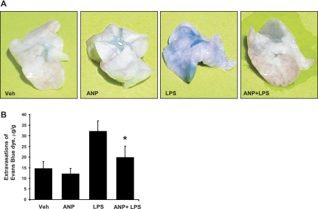
Increased levels of atrial natriuretic peptide (ANP) in the models of sepsis, pulmonary edema, and acute respiratory distress syndrome (ARDS) suggest its potential role in the modulation of acute lung injury. We have recently described ANP-protective effects against thrombin-induced barrier dysfunction in pulmonary endothelial cells (EC). The current study examined involvement of the Rac effector p21-activated kinase (PAK1) in ANP-protective effects in the model of lung vascular permeability induced by bacterial wall LPS. C57BL/6J mice or ANP knockout mice (Nppa(-/-)) were treated with LPS (0.63 mg/kg intratracheal) with or without ANP (2 μg/kg iv). Lung injury was monitored by measurements of bronchoalveolar lavage protein content, cell count, Evans blue extravasation, and lung histology. Endothelial barrier properties were assessed by morphological analysis and measurements of transendothelial electrical resistance. ANP treatment stimulated Rac-dependent PAK1 phosphorylation, attenuated endothelial permeability caused by LPS, TNF-α, and IL-6, decreased LPS-induced cell and protein accumulation in bronchoalveolar lavage fluid, and suppressed Evans blue extravasation in the murine model of acute lung injury. More severe LPS-induced lung injury and vascular leak were observed in ANP knockout mice. In rescue experiments, ANP injection significantly reduced lung injury in Nppa(-/-) mice caused by LPS. Molecular inhibition of PAK1 suppressed the protective effects of ANP treatment against LPS-induced lung injury and endothelial barrier dysfunction. This study shows that the protective effects of ANP against LPS-induced vascular leak are mediated at least in part by PAK1-dependent signaling leading to EC barrier enhancement. Our data suggest a direct role for ANP in endothelial barrier regulation via modulation of small GTPase signaling.


Figures

Fig. 1.
Fig. 1.
Effects of atrial natriuretic peptide (ANP) on endothelial cell (EC) barrier dysfunction induced by inflammatory agonists. EC were plated on gold microelectrodes and grown to confluence. At the time point indicated by the 1st arrow, cells were pretreated with ANP (100 nM, 20 min). At the time point indicated by the 2nd arrow, cells were stimulated with LPS (200 ng/ml; A and B), TNF-α (20 ng/ml; C), or a combination of IL-6 (25 ng/ml) and its soluble coreceptor, IL-6+SR (100 ng/ml; D), and transendothelial electrical resistance was monitored over time. Data are expressed as means ± SD of 3–10 independent experiments; n = 3 per condition for each experiment. HPAEC, human pulmonary macrovascular EC; HLMVEC, human lung microvascular EC.
Fig. 2.
Fig. 2.
Analysis of p21-activated kinase (PAK1) phosphorylation (p-) in response to ANP. HPAEC (top) or HLMVEC (bottom) were treated with either vehicle or ANP (100 nM) for 0.5, 3, 6, or 18 h. Phosphorylation of PAK was detected by Western blot with corresponding phosphospecific antibodies. Equal protein loading was confirmed by determination of PAK1 and β-tubulin content in total cell lysates. Results are representative of 4 independent experiments.
Fig. 3.
Fig. 3.
Effects of ANP on pulmonary vascular leak and inflammation in vivo. Mice were treated with LPS [0.63 mg/kg intratracheal (it)] or vehicle with or without concurrent ANP injection (2 μg/kg iv) for 16 h. Cell counts (A), myeloperoxidase (MPO) activity (B), and protein concentration (C) were determined in bronchoalveolar lavage (BAL) fluid taken from control (Con) and experimental animals (n = 6 per group); *P < 0.001. D: whole lungs were fixed in 10% formaldehyde, embedded in paraffin, and used for histological evaluation by hematoxylin and eosin staining as described in materials and methods (magnification: ×40; n = 3–4 per group, ≥20 fields per slide were inspected).
Fig. 4.
Fig. 4.
Effect of ANP on lung vascular leak induced by LPS. Mice were treated with LPS (0.63 mg/kg it) or vehicle (Veh) with or without concurrent ANP injection (2 μg/kg iv) for 16 h. Effects of ANP on the LPS-induced vascular leak were assessed by measurements of Evans blue leakage into the lung tissue (A). The quantitative analysis of Evans blue-labeled albumin extravasation was performed by spectrophotometric analysis of Evans blue extracted from the lung tissue samples as described in materials and methods (B); n = 6 for each experimental group; *P < 0.01 vs. LPS alone.
Fig. 5.
Fig. 5.
Analysis of LPS-induced ANP expression and PAK phosphorylation in the lungs. Mice were treated with LPS (0.63 mg/kg it) or vehicle for various time points. A: expression of ANP mRNA was determined by quantitative (q) RT-PCR analysis of right and left lung tissue samples. The graph represents pooled data; n = 6 for each experimental group; *P < 0.05. B: PAK phosphorylation in lungs was analyzed by Western blot with phosphospecific antibodies. Equal protein loading was confirmed by probing of membranes with total PAK1 and β-tubulin antibodies. Results of densitometry are shown as means ± SD; n = 6 per group; *P < 0.05. RDU, relative density units.
Fig. 6.
Fig. 6.
Analysis of LPS-induced lung injury in ANP knockout mice. ANP knockout mice (Nppa−/−) or matched controls were treated with LPS (0.63 mg/kg it) for 16 h. A: cell count and protein concentration were measured in BAL fluid as described in materials and methods. B: LPS-induced vascular leak was analyzed by spectrometric determination of Evans blue-labeled albumin extravasation into the lung tissue. C: LPS-induced neutrophilic infiltration was evaluated by histological analysis of lung sections from control and Nppa−/− mice. D: PAK phosphorylation in lung samples from LPS-treated mice was analyzed after 1 h of LPS administration by Western blot with phosphospecific antibodies. Equal protein loading was confirmed by probing of membranes with total PAK1 and β-tubulin antibodies. Results of densitometry are shown as means ± SD; n = 6 per group; *P < 0.05. E and F: effects of single ANP injection (2 μg/kg iv) on LPS-induced lung injury in Nppa−/− mice were assessed by measurements of cell count and protein concentration in the BAL fluid (E) and by analysis of Evans blue accumulation in the lung tissue (F). n = 4–7 per group; *P < 0.05. WT, wild-type.
Fig. 7.
Fig. 7.
Effects of PAK1 depletion on the ANP-mediated barrier protection in human pulmonary EC. EC grown on glass coverslips were transfected with nonspecific (ns) RNA or with PAK1-specific (siPAK1) small interfering RNA (siRNA). A: cells were treated with ANP (100 nM, 20 min) before TNF-α (20 ng/ml, 6 h) challenge. Analysis of cytoskeletal remodeling was performed by double immunofluorescence staining with VE-cadherin and Texas red-phalloidin. Paracellular gaps are marked by arrows. Bottom shows merged images of F-actin and VE-cadherin staining. B: in other experiments, cells were pretreated with ANP (100 nM, 20 min) followed by LPS (200 ng/ml, 6 h). Actin rearrangement was analyzed by immunofluorescence staining for F-actin. C: transendothelial resistance was monitored in control and PAK1-depleted EC over 10 h. Data are expressed as means ± SD of 3–8 independent experiments, n = 3 per experimental condition. Dotted lines mark the time points used for analysis of EC cytoskeletal remodeling depicted in A and B.
Fig. 8.
Fig. 8.
Role of PAK1 in ANP-mediated protection against LPS-induced vascular leak in vivo. Mice were transfected with nonspecific or PAK1-specific siRNA for 72 h. A: Western blot analysis of siRNA-mediated PAK1 knockdown was performed in the lung, heart, liver, and kidney tissue samples. Results of densitometry are shown as means ± SD; *P < 0.05. B: expression of PAK1 mRNA was determined by qRT-PCR analysis of lung tissue samples from control and siPAK1-treated mice (n = 4–6 per condition). C: animals were treated with LPS (0.63 mg/kg it, 16 h) with or without ANP treatment (2 μg/kg iv). After 16 h, cell count and protein concentration were determined in BAL fluid (n = 5–9 per group; *P < 0.001; **P < 0.003). D: whole lungs were used for histological evaluation by hematoxylin and eosin staining as described in materials and methods (magnification: ×40; n = 4 per group, ≥20 fields per slide were inspected). E: effect of ANP on LPS-induced vascular leak was analyzed by Evans blue-labeled albumin extravasation into the lung tissue; n = 5–6 for each experimental group; *P < 0.02.

References

    1. Ahluwalia A, MacAllister RJ, Hobbs AJ. Vascular actions of natriuretic peptides. Cyclic GMP-dependent and -independent mechanisms. Basic Res Cardiol 99: 83–89, 2004 - PubMed
    1. Anand-Srivastava MB. Natriuretic peptide receptor-C signaling and regulation. Peptides 26: 1044–1059, 2005 - PubMed
    1. Baxter GF. The natriuretic peptides. Basic Res Cardiol 99: 71–75, 2004 - PubMed
    1. Birukov KG, Bochkov VN, Birukova AA, Kawkitinarong K, Rios A, Leitner A, Verin AD, Bokoch GM, Leitinger N, Garcia JG. Epoxycyclopentenone-containing oxidized phospholipids restore endothelial barrier function via Cdc42 and Rac. Circ Res 95: 892–901, 2004 - PubMed
    1. Birukova AA, Alekseeva E, Cokic I, Turner CE, Birukov KG. Cross talk between paxillin and Rac is critical for mediation of barrier-protective effects by oxidized phospholipids. Am J Physiol Lung Cell Mol Physiol 295: L593–L602, 2008 - PMC - PubMed

Publication types

MeSH terms