. 2010 Oct 22;285(43):32999-33009.
doi: 10.1074/jbc.M110.108175. Epub 2010 Aug 21.

Angiopoietin-like 4 interacts with matrix proteins to modulate wound healing

Affiliations

Angiopoietin-like 4 interacts with matrix proteins to modulate wound healing

Yan Yih Goh et al. J Biol Chem. .

Abstract

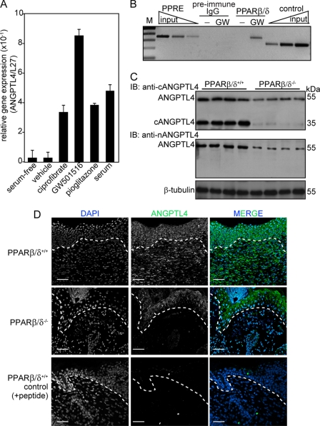
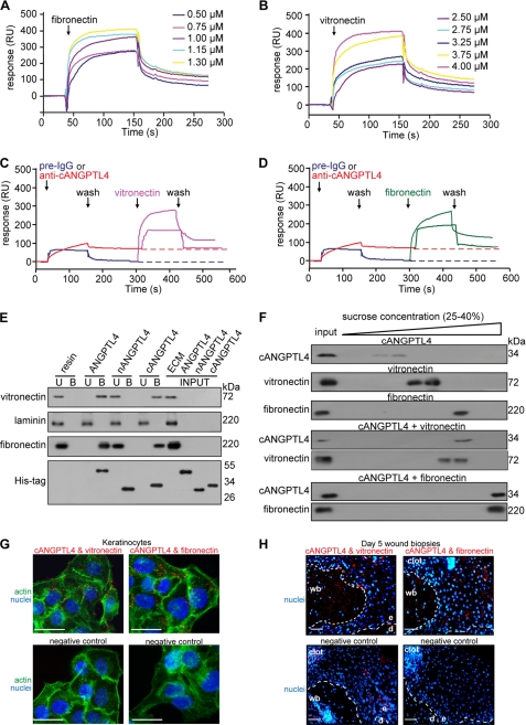
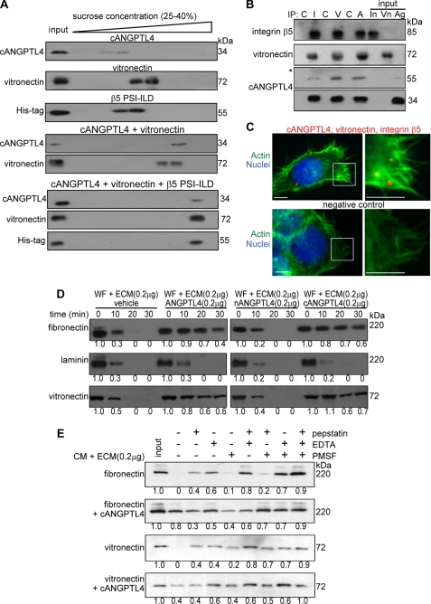
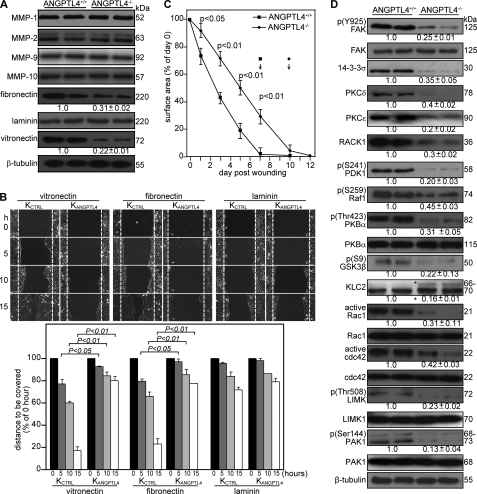
A dynamic cell-matrix interaction is crucial for a rapid cellular response to changes in the environment. Appropriate cell behavior in response to the changing wound environment is required for efficient wound closure. However, the way in which wound keratinocytes modify the wound environment to coordinate with such cellular responses remains less studied. We demonstrated that angiopoietin-like 4 (ANGPTL4) produced by wound keratinocytes coordinates cell-matrix communication. ANGPTL4 interacts with vitronectin and fibronectin in the wound bed, delaying their proteolytic degradation by metalloproteinases. This interaction does not interfere with integrin-matrix protein recognition and directly affects cell-matrix communication by altering the availability of intact matrix proteins. These interactions stimulate integrin- focal adhesion kinase, 14-3-3, and PKC-mediated signaling pathways essential for effective wound healing. The deficiency of ANGPTL4 in mice delays wound re-epithelialization. Further analysis revealed that cell migration was impaired in the ANGPTL4-deficient keratinocytes. Altogether, the findings provide molecular insight into a novel control of wound healing via ANGPTL4-dependent regulation of cell-matrix communication. Given the known role of ANGPTL4 in glucose and lipid homeostasis, it is a prime therapeutic candidate for the treatment of diabetic wounds. It also underscores the importance of cell-matrix communication during angiogenesis and cancer metastasis.


Figures

FIGURE 1.
FIGURE 1.
Reduced expression of ANGPLT4 in PPARβ/δ−/− mice wounds. A, relative mRNA levels of ANGPTL4 in human keratinocytes treated with different agonists selective for each PPAR isotype, ciprofibrate (30 μm, PPARα), GW501516 (2 nm, GW, PPARβ/δ), and pioglitazone (500 nm, PPARγ) are shown. Values are mean ± S.E. of four independent studies. Ribosomal protein L27 used as a normalizing housekeeping gene. B, ChIP was done in keratinocytes using pre-immune IgG or monoclonal anti-PPARβ/δ. Regulatory region with the PPAR response element was immunoprecipitated with anti-PPARβ/δ and specifically amplified. No amplified signal was obtained with preimmune IgG. A control region upstream of PPAR response element served as the negative control. Aliquots of chromatin were analyzed before immunoprecipitation (input). M, 100-bp DNA marker. C, expression is shown of ANGPTL4 protein in day 3 post-wounding mice skin biopsies. Polyclonal antibodies that recognized the N-terminal (anti-nANGPTL4) and C-terminal (anti-cANGPTL4) of ANGPTL4 were used. β-Tubulin served as loading and transfer control. n = 5. IB, immunoblot. D, shown is immunofluorescence staining of ANGPTL4 in PPARβ/δ+/+ and PPARβ/δ−/− day 3 wound biopsies using anti-cANGPTL4. Sections were counterstained with DAPI. Negative control is performed with anti-cANGPTL4 preincubated with antigen peptide. Representative images from wound epithelia and wound beds were shown (n = 5). The dotted white line denotes epidermal-dermal junction. Scale bar, 40 μm.
FIGURE 2.
FIGURE 2.
cANGPTL4 interacts with vitronectin and fibronectin. Sensograms show binding profiles between immobilized-cANGPTL4 and the indicated concentrations of either fibronectin (A) or vitronectin (B). A representative sensorgram (n = 5) shows binding profiles of vitronectin (C, pink) and fibronectin (D, green) with immobilized-cANGPTL4 CM5 chip after preblocking with either pre-immune IgG (blue) or anti-cANGPTL4 (red). E, co-immunoprecipitation assays (n = 5) were carried out by using different forms of His-tagged ANGPTL4 proteins immobilized on nickel-Sepharose and incubated with the indicated purified matrix molecules. Matrix proteins and ANGPTL4 were detected by immunodetected using corresponding antibodies and revealed by chemiluminescence. U and B denote the unbound/washed and bound fractions from the resin, respectively. RU, resonance units. F, shown is immunodetection of the indicated proteins from sucrose density gradient fractions. The proteins were allowed to interact in the indicated combinations before separation by sucrose gradient ultracentrifugation. Blots showed increasing sucrose density from left to right. An aliquot of the indicated protein, prior incubation, and centrifugation was also loaded (input) (n = 5). Detection of various complexes between ANGPTL4 and the indicated binding partners in KCTRL (G) and in Day-5 (H) wound biopsies using PLA is shown. PLA signals (red), nuclei stained with Hoechst dye (blue), and actin stress fiber (green) by Alexa488-phalloidin is shown. The nuclei-image has been acquired in one z-plane using LSM710 confocal microscope. The dotted white line represents epidermal-dermal junction. Negative control is performed without primary antibodies. Representative pictures from wound section with epidermis (e), dermis (d), the adjacent wound bed (wb), and KCTRL from six independent experiments or sections from three mice are shown. Scale bar, 40 μm.
FIGURE 3.
FIGURE 3.
ANGPTL4 modulates matrix protein degradation. A, shown are immunoblots of cANGPTL4, His-tagged integrin β5 PSI-ILD, and vitronectin of sucrose density gradient fractions. The proteins were allowed to interact in the indicated combinations before separation by sucrose density gradient ultracentrifugation. Blots showed increasing sucrose density from left to right. B, in vivo co-immunoprecipitation (IP; n = 3) was performed by using antibodies against integrin β5 (I), vitronectin (V), and cANGPTL4 (A). As control, pre-immune IgG (C) was used. Antibodies were covalently cross-linked to Protein G on agarose beads were incubated with total keratinocyte cell lysate. Immunoprecipitates were detected by immunoblot using corresponding antibodies and revealed by chemiluminescence. Native ANGPTL4 of ∼55 kDa was after longer exposure time as denoted by asterisk. Total cell lysate served as the input. (In, integrin β5; Vn, vitronectin; Ag, ANGPTL4). C, a triple PLA showed the ternary complex in keratinocytes. Triple PLA signals (red) nuclei were stained with Hoechst dye (blue) and Alexa488-phallodin for actin fiber (green). Representative PLA images from three independent experiments are shown. Negative control is without anti-cANGPLT4 and anti-integrin proximity probes. Scale bar, 40 μm. Immunodetection of the matrix proteins, vitronectin, and fibronectin after incubation for indicated time with WF (D) or TNF-α-treated KANGPTL4 (E) CM in the presence of indicated protease inhibitors is shown. Laminin, which does not interact with cANGPTL4, serves as control. Three independent experiments from two wound fluids were performed. Values below denote change in mean -fold expression compared with input.
FIGURE 4.
FIGURE 4.
ANGPTL4 knock-out mice displayed impaired wound re-epithelialization. A, shown is an immunoblot analysis of MMP-1, -2, -9, -10, fibronectin, laminins, and vitronectin from day 5 wound biopsies of ANGPTL4+/+ and ANGPTL4−/− mice. Values below the band represent the mean -fold differences in protein expression levels relative to ANGPTL4+/+ from eight wound biopsies for each genotype. β-Tubulin served as loading and transfer controls. B, shown are wound closure kinetics of KCTRL and KANGPTL4 treated with mitomycin C (2 μg/ml) on the indicated matrix protein-coated surfaces. Representative time-lapsed images of wounded cultures are shown. Yellow dotted lines represent the scratch gap at the time of wounding. The graph shows the distance to be covered by the migrating keratinocytes as the percentage of 0 h (=100%) in vitro wound gap distance (±S.E., n = 5, using the Mann-Whitney test). C, shown are the wound closure kinetics of ANGPTL4+/+ and ANGPTL4−/− mice. Wound surface areas are plotted as percentage of day 0 (=100%) wound surface area (±S.E., n = 10, using the Mann-Whitney test). Arrows indicate the mean time for complete wound closure. D, shown are immunoblot analyses of the indicated proteins from ANGPTL4+/+ and ANGPTL4−/− mice day 5 wound biopsies (n = 5). Values below the bands represent the mean -fold differences in protein expression levels when compared with ANGPTL4+/+, which was assigned the value 1. GSK-3, glycogen synthase kinase-3; KLC, kinesin-light chain; PAK, p21-activated kinase; PDK1, 3-phosphoinositide-dependent kinase-1; LIMK, LIM kinase. β-Tubulin served as the loading and transfer control.

Similar articles

Cited by

References

    1. Hynes R. O. (2002) Cell 110, 673–687 - PubMed
    1. Werner S., Grose R. (2003) Physiol. Rev. 83, 835–870 - PubMed
    1. Bornstein P., Sage E. H. (2002) Curr. Opin Cell Biol. 14, 608–616 - PubMed
    1. Bulcão C., Ferreira S. R., Giuffrida F. M., Ribeiro-Filho F. F. (2006) Curr. Diabetes Rev. 2, 19–28 - PubMed
    1. Caswell P. T., Norman J. C. (2006) Traffic 7, 14–21 - PubMed

Publication types

MeSH terms