. 2010 Oct 29;285(44):34115-25.
doi: 10.1074/jbc.M110.120659. Epub 2010 Aug 21.

Nuclear factor I-C regulates TGF-{beta}-dependent hair follicle cycling

Affiliations

Nuclear factor I-C regulates TGF-{beta}-dependent hair follicle cycling

Genta Plasari et al. J Biol Chem. .

Abstract

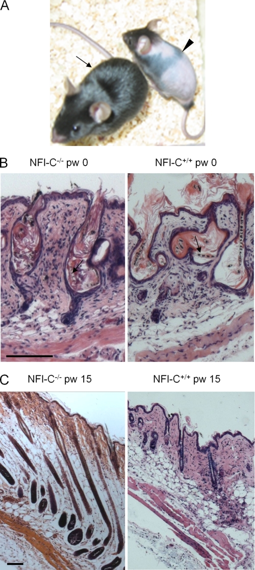
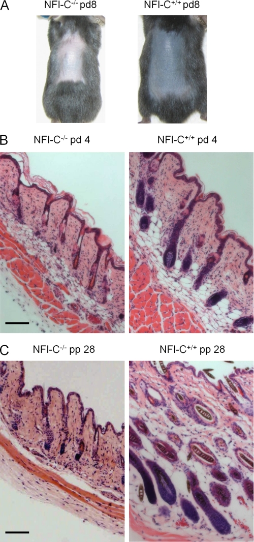
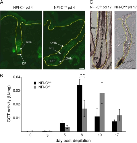
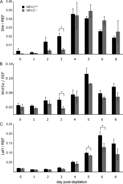
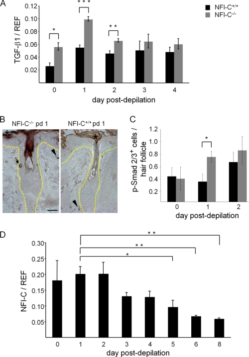
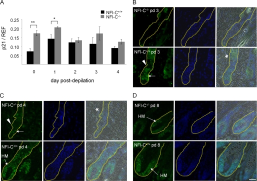
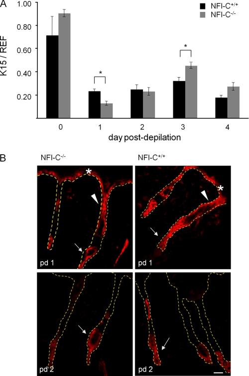
Skin appendages such as teeth and hair share several common signaling pathways. The nuclear factor I C (NFI-C) transcription factor has been implicated in tooth development, but a potential role in hair growth had not been assessed. In this study we found that NFI-C regulates the onset of the hair growth cycle. NFI-C(-/-) mice were delayed in the transition from the telogen to anagen phase of the hair follicle cycle after either experimental depilation or spontaneous hair loss. Lack of NFI-C resulted in delayed induction of the sonic hedgehog, Wnt5a, and Lef1 gene expression, which are key regulators of the hair follicle growth initiation. NFI-C(-/-) mice also showed elevated levels of transforming growth factor β1 (TGF-β1), an inhibitor of keratinocyte proliferation, and of the cell cycle inhibitor p21 at telogen. Reduced expression of Ki67, a marker of cell proliferation, was noted at the onset of anagen, indicating impaired activation of the hair progenitor cells. These findings implicate NFI-C in the repression of TGF-β1 signaling during telogen stage, resulting in the delay of progenitor cell proliferation and hair follicle regeneration in NFI-C-deficient mice. Taken together with prior observations, these findings also designate NFI-C as a regulator of adult progenitor cell proliferation and of postnatal tissue growth or regeneration.


Figures

FIGURE 1.
FIGURE 1.
Hair growth delay phenotype identified in over-groomed NFI-C−/− mice. A, observation of the hair growth 10 days after weaning in an over-groomed litter is shown. The wild-type mouse recovered its normal fur (arrow), whereas the knock-out animal still exhibited a hairless body (arrowhead). B, hematoxylin eosin staining of skin sections performed at weaning (pw 0) and 15 days post-weaning (pw 15) are shown. Arrows indicate the broken hair shafts within the follicle epithelium of telogen phase hair follicles from sections of wild-type and knock-out mice at weaning, probably as a result of the repetitive licking of the fur and skin. C, representative illustrations of normal hair follicle structures 15 days after weaning are shown. Note the telogen phase wild-type hair follicles, as characterized by thin follicles, whereas those of NFI-C-null mice were still in anagen. Scale bars: 100 μm.
FIGURE 2.
FIGURE 2.
NFI-C is involved in the hair follicle cycling. A, 8 days after hair depilation, hair growth was delayed in NFI-C−/− mice, as evidenced by the difference of the back skin color. Note the gray-colored skin of wild-type mice typical of anagen phase pigmentation, whereas the knock-out mice still displayed a pink-colored skin. B, 4 days after depilation (pd 4), the hematoxylin eosin staining of skin histological sections reveals wild-type follicles in early anagen, as indicated by their location at the border between the dermis and the subcutis and the increased size of dermal papillae. In contrast, mutant follicles are just entering anagen and are still located in the dermis. C, shown is hematoxylin eosin staining of histological sections performed at day 28 post-partum (pp 28) of skins not submitted to over-grooming or depilation. Progression to anagen during spontaneous activation of the follicle cycle is delayed in knock-out versus normal animals, as evidenced by the different structure and location of hair follicles. Whereas wild-type animals progressed to the anagen phase, mutant mice are still in telogen. Scale bars: 100 μm.
FIGURE 3.
FIGURE 3.
NFI-C-null mice have delayed anagen onset but otherwise normal hair follicle cycle progression. Molecular markers of the hair follicle cycle progression were analyzed in histological sections of skin collected along the cycle at the indicated days post-depilation (pd). A, 4 days after depilation, the wild-type follicles exhibited a staining typical of the anagen III stage, with P-cadherin being expressed in ORS and matrix but not in the IRS. The knock-out follicle showed a staining typical of the telogen phase as only the secondary hair germ exhibits a strong staining. B, γ-glutamyl transpeptidase colorimetric assay was carried on extracts of skins collected at various times after depilation to encompass the first hair follicle cycle of normal and mutated animals. The error bars represent S.E.; **, indicates p < 0.01. C, staining for the alkaline phosphatase activity was similar in the dermal papilla (DP) of knock-out and wild-type 17 days after plucking. Knock-out animals displayed an additional staining in the ORS characteristic of the end of catagen, whereas wild-type follicles entering telogen do not. The yellow dashed lines indicate the limit of keratinocyte epidermal layers determined by contrast phase. DHM, developing hair matrix; SHG, secondary hair germ. Scale bars: 20 μm.
FIGURE 4.
FIGURE 4.
NFI-C−/− mice display altered signaling pathways controlling anagen onset. The expression levels of Shh (panel A), Wnt5a (panel B), and Lef1 (panel C) relative to reference (REF) genes were assayed by RT-qPCR performed on extract from skin biopsies collected to the indicated time points after depilation. *, indicates p < 0.05.
FIGURE 5.
FIGURE 5.
NFI-C controls TGF-β1 levels during telogen phase. A, TGF-β1 expression relative to reference (REF) genes assayed on total skin extract by RT-qPCR at the indicated days after the depilation. B, immunostaining of p-Smad2/3 at day 1 post-depilation. A positive cell in the ORS of knock-out hair follicle is indicated by an arrow, whereas no staining was detected in wild-type follicular keratinocytes. Arrowheads indicate positive dermal fibroblasts. A nonspecific brown staining was detected in the corneal layers (asterisks). The yellow dashed lines indicate the limit of the keratinocyte epidermal layer. Scale bar: 25 μm. C, the average number ± S.E. of p-Smad2/3-positive cells per hair follicle was calculated in knock-out and wild-type mice at day 0 to day 2 post-depilation. D, NFI-C mRNA expression was assessed by the RT-qPCR assay of extracts of skin biopsies from wild-type animals collected at the indicated time points after depilation. NFI-C expression levels were significantly more elevated during telogen or the telogen-anagen transition (days 0–2) as compared with anagen (days 5–8). *, p < 0.05; **, p < 0.01; ***, p < 0.001.
FIGURE 6.
FIGURE 6.
NFI-C regulates the expression of cell cycle inhibitor p21 and the activation of keratinocyte proliferation at anagen onset. A, p21 mRNA levels were analyzed by RT-qPCR in samples of skin collected from day 0 to day 4 after depilation and they were normalized to reference (REF) mRNAs. A significant increase of p21 was observed at days 0 and 1 in knock-out mice compared with wild-type littermates (*, p < 0.05; **, p < 0.01). B–D, keratinocytes were labeled for Ki67 antigen at day 3 (B), day 4 (C), and day 8 (D) post-depilation (pd). Nuclei were stained with an antibody against Ki67 (green) and DAPI (blue), and the phase contrast, Ki67, and DAPI signals are merged. The asterisks show the location of the arrector pili muscle, whereas the yellow dashed lines indicate the limit of keratinocyte layer. At days 3 and 4, proliferating cells of the secondary hair germ positive for Ki67 are indicated by arrows, whereas the Ki67-negative slow cycling cells of the bulge were shown by the arrowhead. No proliferative signal was observed at day 3, and a weak signal was detected at day 4 in mutant mice. At days 4 and 8, keratinocytes that proliferate in the hair matrix (HM) to form the new shaft are indicated by the HM-labeled arrows. Scale bar: 25 μm.
FIGURE 7.
FIGURE 7.
TGF-β1 levels correlate to the amount of K15-positive stem cells. A, expression of K15 relative to reference (REF) genes assessed in skin samples by RT-qPCR, decreased at day 1 post-depilation. Different levels of expression were observed in wild-type and knock-out samples at days 1 and 3 (*, p < 0.05). B, immunofluorescent staining on skin cryosections localized K15 in the basal layer of the epidermis (asterisks), in the ORS (arrowheads), and in the region corresponding to the bulge and/or second hair germ (arrows) at day 1 post-depilation (pd 1). At day 2 post-depilation the staining was limited to the bulge/second hair germ area (arrows). No significant difference was seen when comparing cell staining of the two genotypes. Yellow dashed lines indicate the limit of keratinocyte layers in the epidermis and hair follicles. Scale bar: 30 μm.

Similar articles

Cited by

References

    1. Gronostajski R. M., Adhya S., Nagata K., Guggenheimer R. A., Hurwitz J. (1985) Mol. Cell. Biol. 5, 964–971 - PMC - PubMed
    1. Roulet E., Bucher P., Schneider R., Wingender E., Dusserre Y., Werner T., Mermod N. (2000) J. Mol. Biol. 297, 833–848 - PubMed
    1. Gronostajski R. M. (2000) Gene 249, 31–45 - PubMed
    1. das Neves L., Duchala C. S., Tolentino-Silva F., Haxhiu M. A., Colmenares C., Macklin W. B., Campbell C. E., Butz K. G., Gronostajski R. M., Godinho F. (1999) Proc. Natl. Acad. Sci. U.S.A. 96, 11946–11951 - PMC - PubMed
    1. Gründer A., Ebel T. T., Mallo M., Schwarzkopf G., Shimizu T., Sippel A. E., Schrewe H. (2002) Mech. Dev. 112, 69–77 - PubMed

Publication types