. 2010 Oct 29;285(44):33875-84.
doi: 10.1074/jbc.M110.147264. Epub 2010 Aug 21.

Lipid binding requirements for oxysterol-binding protein Kes1 inhibition of autophagy and endosome-trans-Golgi trafficking pathways

Affiliations

Lipid binding requirements for oxysterol-binding protein Kes1 inhibition of autophagy and endosome-trans-Golgi trafficking pathways

Marissa A LeBlanc et al. J Biol Chem. .

Abstract

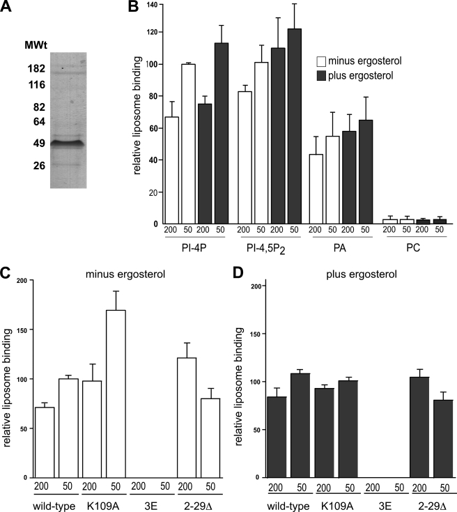
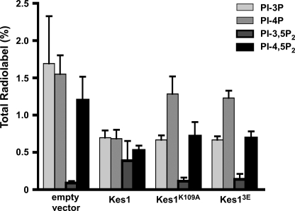
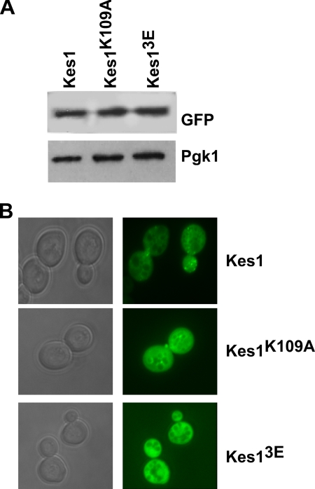
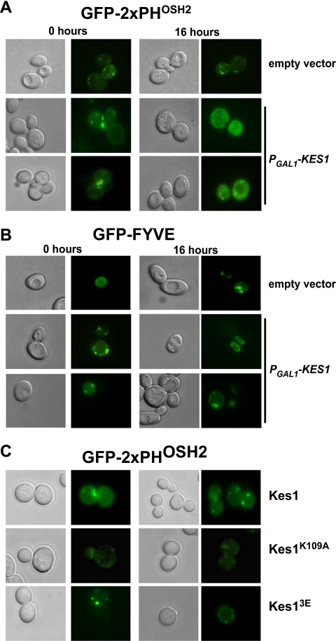
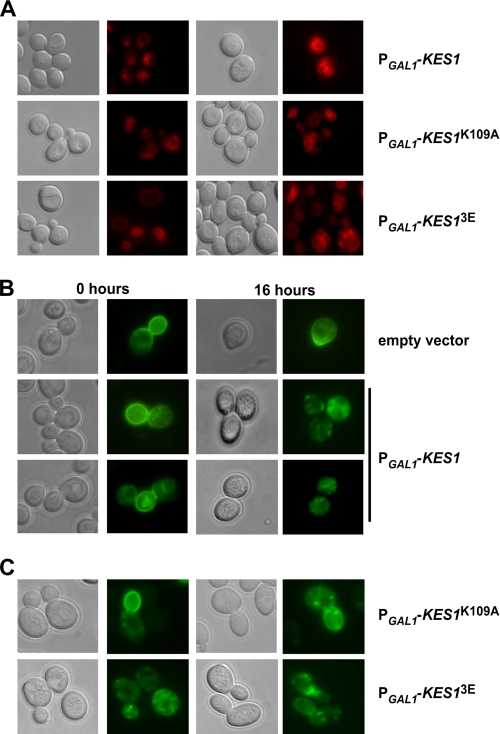
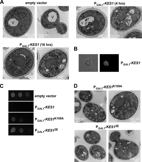
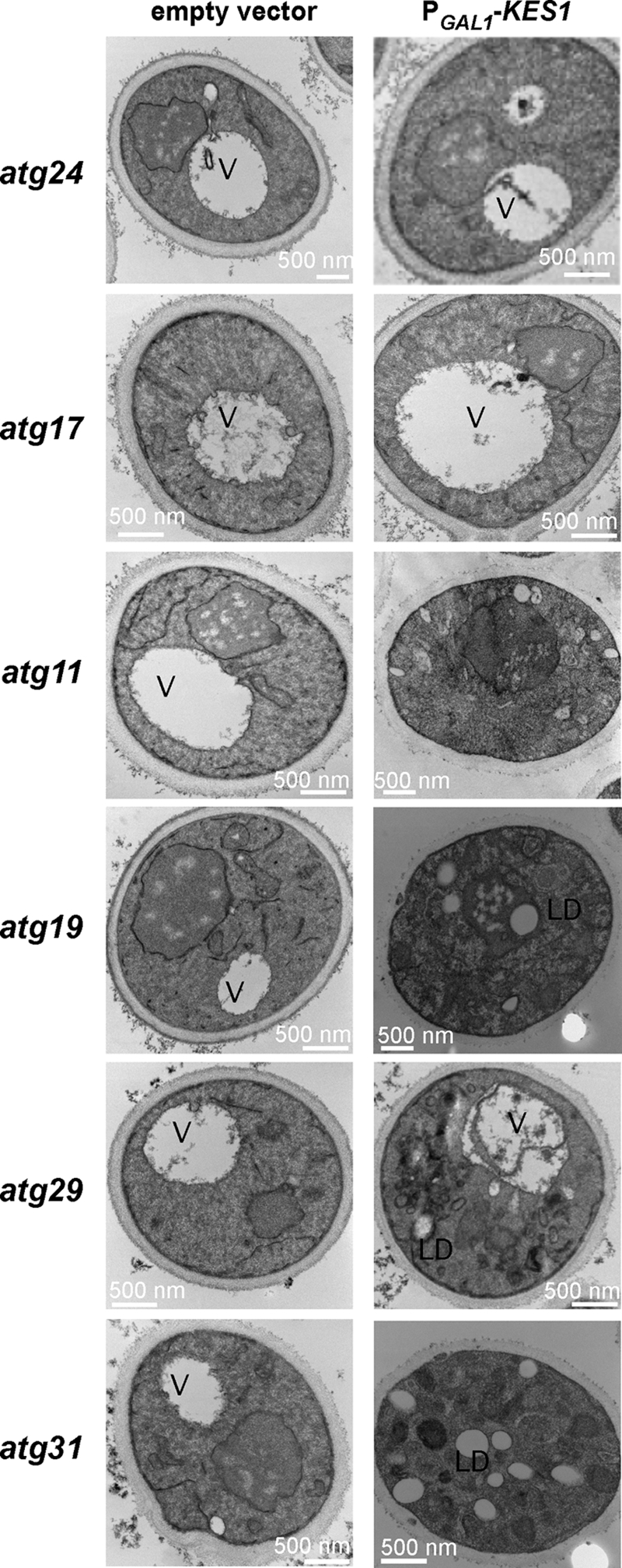
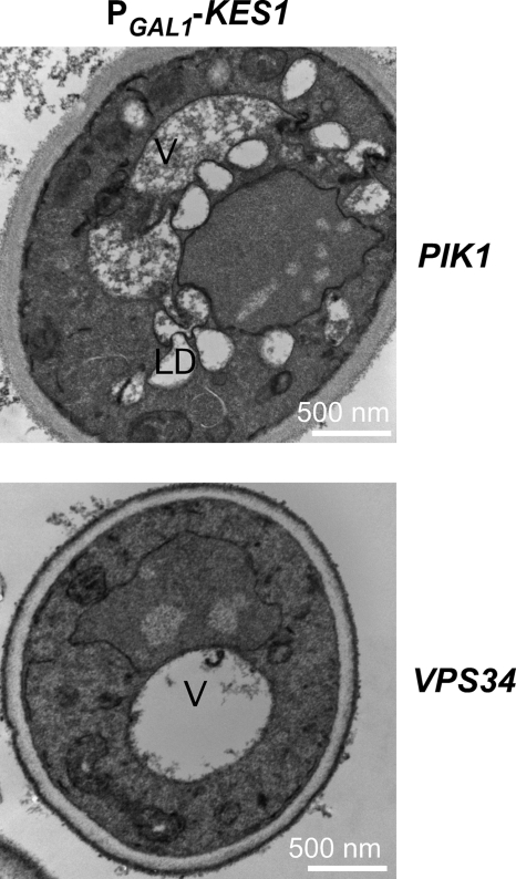
The Saccharomyces cerevisiae protein Kes1/Osh4 is a member of the enigmatic family of oxysterol-binding proteins found throughout Eukarya united by a β-barrel structure that binds sterols and oxysterols. In this study, we determined that phosphoinositides are the major determinant in membranes that facilitate Kes1 association both in vitro and in cells. Increased expression of Kes1 in yeast cells decreased the levels of both phosphatidylinositol 4-phosphate (PI4P) and phosphatidylinositol 3-phosphate (PI3P). Phosphoinositide and sterol bindings by Kes1 were necessary for Kes1 to decrease the level of PI4P but not PI3P. Kes1 inhibited vesicular trafficking between the trans-Golgi and plasma membrane as evidenced by accumulation of the vacuolar soluble NSF attachment protein receptors Snc1 in the cytoplasmic vesicles. Sterol and phosphoinositide binding by Kes1 both contributed to its regulation of Snc1 trafficking. This study also describes a previously unknown role for Kes1 in the regulation of the autophagy/cytoplasm to the vacuole trafficking pathway. The Kes1-mediated regulation of the autophagy/cytoplasm to the vacuole trafficking pathway was prevented by increasing expression of the PI3K Vps34, suggesting that it is the Kes1-mediated decrease in PI3P level that contributes to this regulation.


Figures

FIGURE 1.
FIGURE 1.
Phospholipid with a free phosphate group is required for Kes1 membrane association. A, Kes1-His6 was purified to apparent homogeneity. Purified protein sample was separated by SDS-PAGE, and proteins were stained using GelCode (Pierce). B, liposomes bound to Kes1-His6 were isolated by flotation. Kes1-His6 (0.25 μm) was assayed for binding to liposomes containing PC and 10% PI4P, PI-4,5-P2, or PA (PI3P, PI-3,5-P2, PI, phosphatidylserine, phosphatidylethanolamine, or diacylglycerol, data not shown) ± 10% ergosterol. Kes1-His6 bound liposomes containing all phosphoinositides and PA but not other lipids tested. Mutant Kes1 (Kes1K109A, Kes13E, and Kes12–29Δ) proteins were assayed for binding to liposomes containing PC + 10% PI4P (C), PC + 10% PI4P + 10% ergosterol (D). Assays contained 1 mm liposomes extruded at 200 and 50 nm. After centrifugation, the top and bottom fractions were collected and analyzed by SDS-PAGE and Western blot. The results were quantified using densitometry. Error bars show mean ± S.E. from a minimum of three different experiments using different sets of extruded liposomes and different protein preparations.
FIGURE 2.
FIGURE 2.
Kes1 requires PIP and sterol binding to regulate PI4P level but not PI3P. The kes1Δ strain transformed with the plasmids PGAL1-KES1-His6-pESC-URA, PGAL1-KES1-K109A-His6-pESC-URA, or PGAL1-KES13E-His6-pESC-URA were grown to mid-logarithmic phase at 25 °C. For expression of the KES1 gene from the PGAL1 promoter, 2% galactose was added to the medium for 4 h. Cells were washed in inositol-free medium and labeled with myo-[3H]inositol for 1 h. Phosphoinositides were extracted, deacylated, separated by high performance liquid chromatography and quantified using an on-line radiometric detector. Data are presented as percentage of the total number of counts/min in inositol-containing phospholipids and are expressed as mean ± S.E. of a minimum of three separate experiments.
FIGURE 3.
FIGURE 3.
Kes1 binding to PIPs is required for membrane association in cells. KES1-GFP, KES1K109A-GFP, and KES13E-GFP are expressed under their own promoter in a low copy plasmid (1–2 copies) in a cell with an inactivated KES1 gene. A, Western blot of Kes1-GFP, Kes1K109A-GFP, and Kes13E-GFP shows equal expression of wild-type and mutant Kes1 proteins. B, cells were visualized by using fluorescence (GFP) and differential interference contrast filters.
FIGURE 4.
FIGURE 4.
Sterol and PIP binding by Kes1 is required for proper PI4P distribution. Wild type BY4741 cells containing the empty vector (pESC-LEU) or the plasmid PGAL1-KES1-pESC-LEU were also transformed with the PI4P reporter (GFP-2xPHOSH2) (A) or the PI3P reporter (FYVE-GFP) (B). Cells were grown to mid-logarithmic phase in raffinose medium and transferred to galactose medium for 16 h. C, kes1Δ strain containing the plasmids KES1-pRS415, KES1K109A-pRS415, or KES13E-pRS415 was transformed with the PI4P reporter (GFP-2×PHOSH2). Cells were visualized by using fluorescence (GFP) and differential interference contrast filters.
FIGURE 5.
FIGURE 5.
Kes1 expression inhibits Snc1-GFP transport. A, kes1Δ strain transformed with the plasmids PGAL1-KES1-His6-pESC-URA, PGAL1-KES1K109A-His6-pESC-URA, or PGAL1-KES13E-His6-pESC-URA were grown to mid-logarithmic phase at 25 °C in raffinose medium and then shifted to galactose-containing medium for 16 h. Cells were labeled for 5 min with 40 μm FM4-64 and incubated post-label in fresh medium containing galactose for the indicated time points. kes1Δ strains containing plasmids PGAL1-KES1-His6-pESC-URA(B) and PGAL1-KES1K109A-His6-pESC-URA or PGAL1-KES13E-His6-pESC-URA (C) were transformed with a plasmid expressing Snc1-GFP. Cells were grown to mid-logarithmic phase at 25 °C in raffinose medium and shifted to galactose medium for 16 h and visualized.
FIGURE 6.
FIGURE 6.
Increased Kes1 causes vacuole fragmentation, lipid droplet formation, and membrane accumulation. A, wild type strain BY4741 was transformed with the empty vector pESC-URA, and the kes1Δ strain was transformed with the plasmid PGAL1-KES1-His6-pESC-URA. Cells were grown to mid-logarithmic phase at 25 °C in raffinose medium and shifted to galactose medium for 4 or 16 h and then processed and visualized by electron microscopy. B, kes1Δ strain transformed with PGAL1-KES1-His6-pESC-URA was grown to mid-logarithmic phase at 25 °C in raffinose medium and shifted to galactose-containing medium for 16 h. Cells were stained with 50 μm BODIPY 493/503 for 30 min, washed with PBS, and visualized using differential contrast and fluorescence microscopy (RFP). C, kes1Δ strain transformed with PGAL1-KES1-His6-pESC-URA, PGAL1-KES1K109A-His6-pESC-URA, or PGAL1-KES13E-His6-pESC-URA were grown to mid-logarithmic phase at 25 °C in raffinose medium and shifted to galactose medium for 16 h. Cells were concentrated to 0.1 absorbance units, and a series of serial dilutions were plated on SGal medium and grown for 3 days at 25 °C. D, kes1Δ strain transformed with PGAL1-KES1K109A-His6-pESC-URA or PGAL1-KES13E-His6-pESC-URA was grown to mid-logarithmic phase at 25 °C in raffinose medium and shifted to galactose medium for 16 h before being prepared for electron microscopy. Abbreviations used are as follows: V, vacuole; LD, lipid droplet.
FIGURE 7.
FIGURE 7.
Kes1 causes membrane and lipid droplet accumulation. The empty vector pESC-URA or the plasmid PGAL1-KES1-His6-pESC-URA was transformed into yeast strains with inactivated genes specific for autophagy (atg17Δ, atg29Δ, and atg31Δ) or the Cvt pathway (atg11Δ, atg19Δ, and atg24Δ). Cells were grown to mid-logarithmic phase at 25 °C in raffinose medium and shifted to galactose medium for 16 h prior to visualization by electron microscopy. Abbreviations used are as follows: V, vacuole; LD, lipid droplet.
FIGURE 8.
FIGURE 8.
Increased expression of the PI3K, Vps34, prevents membrane and accumulation defects associated with expression of KES1. The plasmid PGAL1-KES1-pESC-LEU was transformed into a kes1Δ yeast strain expressing either the PIK1 or VPS34 gene on a high copy plasmid. The yeast strains were grown to mid-logarithmic phase at 25 °C in raffinose medium and shifted to galactose-containing medium for 16 h prior to visualization by electron microscopy. Abbreviations used are as follows: V, vacuole; LD, lipid droplet.

Similar articles

Cited by

References

    1. Fairn G. D., McMaster C. R. (2008) Cell. Mol. Life Sci. 65, 228–236 - PMC - PubMed
    1. Raychauduri J., Prinz W. (2010) Annu. Rev. Cell Dev. Biol., in press - PMC - PubMed
    1. Beh C. T., Cool L., Phillips J., Rine J. (2001) Genetics 157, 1117–1140 - PMC - PubMed
    1. Beh C. T., Rine J. (2004) J. Cell Sci. 117, 2983–2996 - PubMed
    1. Im Y. J., Raychaudhuri S., Prinz W. A., Hurley J. H. (2005) Nature 437, 154–158 - PMC - PubMed

Publication types