

Development of a sphingosylphosphorylcholine detection system using RNA aptamers
- PMID: 20729797
- PMCID: PMC6257670
- DOI: 10.3390/molecules15085742
Development of a sphingosylphosphorylcholine detection system using RNA aptamers
Abstract
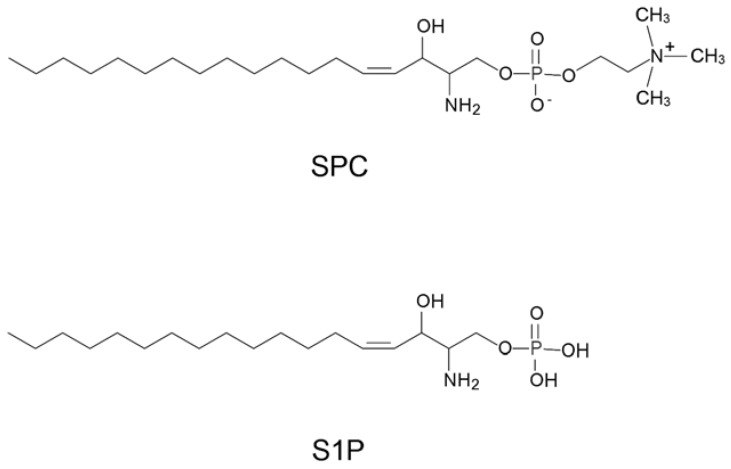
Sphingosylphosphorylcholine (SPC) is a lysosphingolipid that exerts multiple functions, including acting as a spasmogen, as a mitogenic factor for various types of cells, and sometimes as an inflammatory mediator. Currently, liquid chromatography/tandem mass spectrometry (LC/MS/MS) is used for the quantitation of SPC. However, because of the complicated procedures required it may not be cost effective, hampering its regular usage in a routine practical SPC monitoring. In this report, we have generated RNA aptamers that bind to SPC with high affinity using an in vitro selection procedure and developed an enzyme-linked aptamer assay system using the minimized SPC aptamer that can successfully distinguish SPC from the structurally related sphingosine 1-phosphate (S1P). This is the first case of the Systematic Evolution of Ligands by EXponential enrichment (SELEX) process being performed with a lysosphingolipid. The SPC aptamers would be valuable tools for the development of aptamer-based medical diagnosis and for elucidating the biological role of SPC.
Figures


Similar articles
-
In vitro selection of high-affinity DNA aptamers for streptavidin.Acta Biochim Biophys Sin (Shanghai). 2009 Apr;41(4):335-40. doi: 10.1093/abbs/gmp022. Acta Biochim Biophys Sin (Shanghai). 2009. PMID: 19352549
-
Aptamers: selection, modification and application to nervous system diseases.Curr Med Chem. 2011;18(27):4159-68. doi: 10.2174/092986711797189646. Curr Med Chem. 2011. PMID: 21838689 Review.
-
Development of Cell-Specific Aptamers: Recent Advances and Insight into the Selection Procedures.Molecules. 2017 Nov 27;22(12):2070. doi: 10.3390/molecules22122070. Molecules. 2017. PMID: 29186905 Free PMC article. Review.
-
In vitro selection of RNA aptamers that selectively bind danofloxacin.Biochem Biophys Res Commun. 2014 Jun 13;448(4):397-402. doi: 10.1016/j.bbrc.2014.04.103. Epub 2014 May 2. Biochem Biophys Res Commun. 2014. PMID: 24792181
-
Selection of DNA aptamers that bind to influenza A viruses with high affinity and broad subtype specificity.Biochem Biophys Res Commun. 2014 Jan 3;443(1):37-41. doi: 10.1016/j.bbrc.2013.11.041. Epub 2013 Nov 19. Biochem Biophys Res Commun. 2014. PMID: 24269231
Cited by
-
SELEX tool: a novel and convenient gel-based diffusion method for monitoring of aptamer-target binding.J Biol Eng. 2020 Jan 13;14:1. doi: 10.1186/s13036-019-0223-y. eCollection 2020. J Biol Eng. 2020. PMID: 31956340 Free PMC article.
-
Identification and characterization of a mirror-image oligonucleotide that binds and neutralizes sphingosine 1-phosphate, a central mediator of angiogenesis.Biochem J. 2014 Aug 15;462(1):153-62. doi: 10.1042/BJ20131422. Biochem J. 2014. PMID: 24832383 Free PMC article.
-
NMR monitoring of the SELEX process to confirm enrichment of structured RNA.Sci Rep. 2017 Mar 21;7(1):283. doi: 10.1038/s41598-017-00273-x. Sci Rep. 2017. PMID: 28325909 Free PMC article.
-
Label-Free G-Quadruplex Aptamer Fluorescence Assay for Ochratoxin A Using a Thioflavin T Probe.Toxins (Basel). 2018 May 12;10(5):198. doi: 10.3390/toxins10050198. Toxins (Basel). 2018. PMID: 29757205 Free PMC article.
-
Selection, Characterization and Application of Artificial DNA Aptamer Containing Appended Bases with Sub-nanomolar Affinity for a Salivary Biomarker.Sci Rep. 2017 Mar 3;7:42716. doi: 10.1038/srep42716. Sci Rep. 2017. PMID: 28256555 Free PMC article.
References
-
- Higuchi K., Hara J., Okamoto R., Kawashima M., Imokawa G. The skin of atopic dermatitis patients contains a novel enzyme, glucosylceramide sphingomyelin deacylase, which cleaves the N-acyl linkage of sphingomyelin and glucosylceramide. Biochem. J. 2000;350:747–756. doi: 10.1042/0264-6021:3500747. - DOI - PMC - PubMed
Publication types
MeSH terms
Substances
LinkOut - more resources
Full Text Sources
Other Literature Sources
