. 2010 Sep;42(9):745-50.
doi: 10.1038/ng.643. Epub 2010 Aug 22.

A large and complex structural polymorphism at 16p12.1 underlies microdeletion disease risk

Affiliations

A large and complex structural polymorphism at 16p12.1 underlies microdeletion disease risk

Francesca Antonacci et al. Nat Genet. 2010 Sep.

Abstract

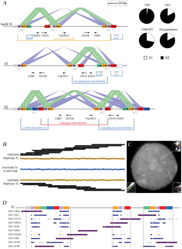
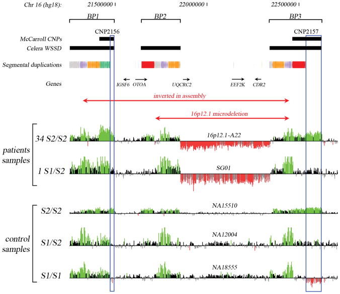
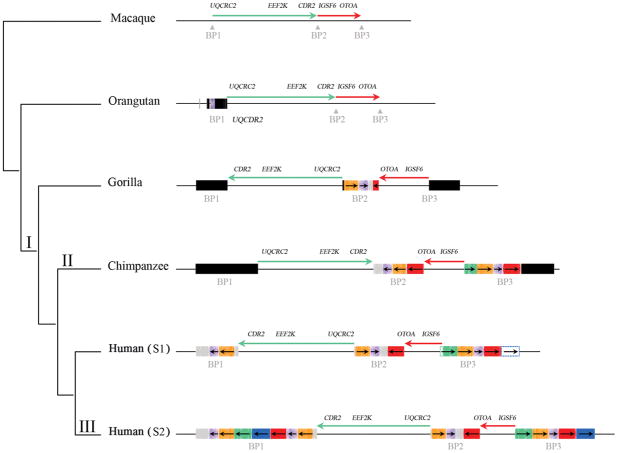
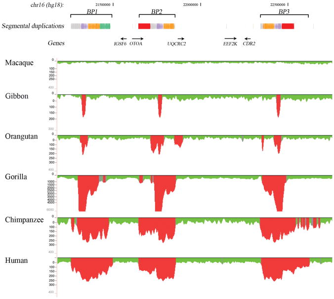
There is a complex relationship between the evolution of segmental duplications and rearrangements associated with human disease. We performed a detailed analysis of one region on chromosome 16p12.1 associated with neurocognitive disease and identified one of the largest structural inconsistencies in the human reference assembly. Various genomic analyses show that all examined humans are homozygously inverted relative to the reference genome for a 1.1-Mb region on 16p12.1. We determined that this assembly discrepancy stems from two common structural configurations with worldwide frequencies of 17.6% (S1) and 82.4% (S2). This polymorphism arose from the rapid integration of segmental duplications, precipitating two local inversions within the human lineage over the last 10 million years. The two human haplotypes differ by 333 kb of additional duplicated sequence present in S2 but not in S1. Notably, we show that the S2 configuration harbors directly oriented duplications, specifically predisposing this chromosome to disease-associated rearrangement.


Conflict of interest statement

FINANCIAL INTEREST

E.E.E. is a member of the Scientific Advisory Board Member of Pacific Biosciences. J.A.R. is employee of Signature Genomic Laboratories, LLC. L.G.S. is an employee of, owns shares in and sits on the Members’ Board of Signature Genomic Laboratories, LLC.

Figures

Figure 1
Figure 1
Alternate structural configurations of the 16p12.1 region. (A) The organization in the reference genome (build 36, top schematic) is compared against two experimentally validated structural configurations (S1 and S2). The locations of the inversion, copy-number polymorphisms (CNP2156 and CNP2157), a rare (20/6712) non-pathogenic deletion variant and segmental duplications (colored rectangles) are indicated. Dashed empty boxes at the S1 structure correspond to regions duplicated in S2 but present in single copy in the S1 haplotype. The S1 and S2 structures differ because of the presence of the distal duplication segment (CNP2156 and CNP2157 at BP1) on the S2 haplotype. Based on this structure, the S1 configuration is predicted to be protective against occurrence of the 16p12.1 pathogenic microdeletion. The red block corresponds to the 68-kbp segmental duplication that likely mediates, through NAHR, the recurrent 16p12.1 microdeletion in patients . Segments duplicated in a direct orientation are connected by green lines while sequences duplicated in an inverted orientation are connected by blue lines. (B) The organization of the region was experimentally validated by optical mapping. SwaI single-molecule restriction maps are depicted and summarized for both configurations (Supplementary Note). (C) The large-scale orientation of each block was confirmed by FISH experiments on interphase nuclei and stretched chromosomes (white rectangles) using probes mapping at the red, blue and green segmental duplications. (D) A contig of 10 BAC clones from the genome of the complete hydatidiform mole (CHM1hTERT) along the 16p12.1 region was sequenced. All clones mapped against the S2 structure were concordant.
Figure 2
Figure 2
ArrayCGH data for 16p12.1 microdeletion patient samples and control HapMap samples (NA15510, NA12004 and NA18555). Probes with log2 ratios above or below a threshold of 1.5 standard deviations from the normalized mean log2 ratio are colored green (duplication) or red (deletion), respectively. The positions of copy-number polymorphisms (CNP2156 and CNP2157) and segmental duplications are indicated. Blue empty boxes highlight the S2-specific duplications that have a diploid copy number of 2 in S1/S1 individuals, 3 in S1/S2 heterozygotes, and 4 in S2/S2 homozygotes. HapMap sample NA18956 with S1/S2 genotype was used as reference.
Figure 3
Figure 3
Expansion and multiple inversions of the 16p12.1 region in humans and the syntenic regions in non-human primates during primate evolution. The genomic organization is compared within a generally accepted phylogeny of macaque, orangutan, gorilla, chimpanzee and human. The region has expanded from 726 kbp (macaque) to 1.6 Mbp (human S2) as a result of segmental duplication accumulation (black and colored rectangles). Sequence and FISH data indicate that the inverted configuration as found in orangutan and macaque is likely the ancestral state in all mammals (I). The expansion of segmental duplications in the African great ape ancestor occurred in conjunction with two inversions between BP1 to BP2 (green arrow) and BP2 to BP3 (red arrow), which may have reverted back to the direct orientation in the chimpanzee lineage (II). The region has become increasingly complex in human leading to the addition of another polymorphic 333 kbp at BP1 specifically in the human lineage (III). Colored boxes indicate segmental duplications as determined by complete sequencing of large-insert BAC clones from primate genomic libraries (Supplementary Note).
Figure 4
Figure 4
Regions of segmental duplication based on read-depth mapping of whole-genome shotgun sequences (WGS) against the human genome. The figure shows an expansion of segmental duplications in the African great apes (human, chimpanzee, gorilla) with respect to orangutan, gibbon and macaque. Also shown are the segmental duplications in human annotated using SegDupMasker .

Comment in

References

    1. Itsara A, et al. Population analysis of large copy number variants and hotspots of human genetic disease. Am J Hum Genet. 2009;84:148–61. - PMC - PubMed
    1. McCarroll SA, et al. Integrated detection and population-genetic analysis of SNPs and copy number variation. Nat Genet. 2008;40:1166–74. - PubMed
    1. Conrad DF, et al. Origins and functional impact of copy number variation in the human genome. Nature. 2009 - PMC - PubMed
    1. Cheung VG, et al. Integration of cytogenetic landmarks into the draft sequence of the human genome. Nature. 2001;409:953–8. - PMC - PubMed
    1. Bailey JA, et al. Recent segmental duplications in the human genome. Science (New York, N Y. 2002;297:1003–7. - PubMed

Publication types