

doi: 10.1038/nm.2199.
Epub 2010 Aug 22.
Inhibitors of leucine-rich repeat kinase-2 protect against models of Parkinson's disease
Affiliations
- PMID: 20729864
- PMCID: PMC2935926
- DOI: 10.1038/nm.2199
Item in Clipboard
Inhibitors of leucine-rich repeat kinase-2 protect against models of Parkinson's disease
Nat Med.
2010 Sep.
Abstract
Leucine-rich repeat kinase-2 (LRRK2) mutations are a common cause of Parkinson's disease. Here we identify inhibitors of LRRK2 kinase that are protective in in vitro and in vivo models of LRRK2-induced neurodegeneration. These results establish that LRRK2-induced degeneration of neurons in vivo is kinase dependent and that LRRK2 kinase inhibition provides a potential new neuroprotective paradigm for the treatment of Parkinson's disease.
Conflict of interest statement
TMD is a paid consultant to Merck KGAA. The terms of this arrangement are being managed by the Johns Hopkins University in accordance with its conflict of interest policies.
Figures

Identification of inhibitors of LRRK2 kinase. (a) LRRK2 autophosphorylation (% of control) ± Biomol inhibitors (See Table S1). Red indicates LRRK2 kinase inhibitors. ***p<0.001 by ANOVA compared to the other groups. Neuman-Keuls post hoc test. Degree of freedom = 34 (total) and F = 18.4144. (b) Representative phosphoimage of WT and LRRK2 G2019S autophosphorylation ± LRRK2 kinase inhibitors. LRRK2 kinase dead (D1994A) and KN-93 are negative controls. (c, d) LRRK2 kinase inhibitors dose-response curves of LRRK2 WT and G2019S autophosphorylation. (e, f, g) Raf kinase inhibitors dose-response curves on LRRK2 WT, LRRK2 G2019S and LRRK1 autophosphorylation. (h) LRRK2 G2019S autophosphorylation and 4E-BP1 phosphorylation ± LRRK2 kinase inhibitors. LRRK2 G2019S kinase dead mutant (G2019S, D1994A), ZM336372 and indirubin are negative controls. (i) Quantification of LRRK2 G2019S autophophorylation and 4E-BP1 phosphorylation ± LRRK2 kinase inhibitors. ***P < 0.001, by ANOVA, Neuman-Keuls post hoc test. Degree of freedom for LRRK2 = 17 (total) and F = 22.401. Degree of freedom for 4E-BP1 = 17 (total) and F = 22.453. All data represents the mean ± S.E.M. from three independent experiments.
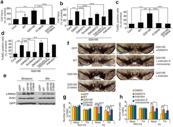
LRRK2 kinase inhibition protects against LRRK2-induced neuronal toxicity. (a) Quantification of neuronal injury, normalized to number of viable neurons transfected with eGFP in three experiments. ***P < 0.001 and *P < 0.05 by ANOVA compared to eGFP control. +++P < 0.001 by ANOVA compared to LRRK2 G2019S. #P < 0.05 by ANOVA compared to LRRK2 D1994A. Tukey-Kramer post hoc test. Degree of freedom = 21 (total) and F = 42.436. (b) Quantification of neuronal injury ± LRRK2 kinase inhibitors. ***P < 0.001 by ANOVA compared to eGFP control. +++P < 0.01 by ANOVA compared to DMSO control. Tukey-Kramer post hoc test. Degree of freedom = 28 (total) and F = 47.3152. (c) Quantification of neuronal cell death via TUNEL. ***P< 0.001 by ANOVA compared to eGFP control. +++P< 0.01 by ANOVA compared to LRRK2 G2019S. Neuman-Keuls post hoc test. Degree of freedom = 14 (total) and F = 12.4378. (d) TUNEL quantification ± LRRK2 kinase inhibitors. **P< 0.01 by ANOVA compared to eGFP control. ++P< 0.01 by ANOVA compared to DMSO control. Neuman-Keuls post hoc test. Degree of freedom = 20 (total) and F = 16.6113. (e) LRRK2 and GFP immunoblots of striatum and substantia nigra (SN) 2 weeks after intrastriatal infusion of HSVPrPUC/CMVeGFP, LRRK2 WT (HSV-LRRK2 WT/CMVeGFP) and LRRK2 G2019S (HSV-LRRK2 G2019S/CMVeGFP). (f) SN tyrosine hydroxylase (TH) immunolabeling 3 weeks after HSV-mediated delivery of eGFP, LRRK2 WT, LRRK2 G2019S, or LRRK2 G2019S, D1994A ± LRRK2 kinase inhibitors. Scale bar = 500 μm. (g) TH-positive and Nissl-positive cell counts comparing eGFP, LRRK2 WT, LRRK2 G2019S, or LRRK2 G2019S, D1994A. Each bar represents the mean number (± S.E.M., n = 8) of TH-positive cells. ***P< 0.001 by ANOVA compared to eGFP control and LRRK2 WT. +++P< 0.001 by ANOVA compared to LRRK2 G2019S, D1994A. Tukey-Kramer post hoc test. Degree of freedom = 67 (total) and F = 6.5115 for Nissl staining groups. Degree of freedom = 68 (total) and F = 7.1292 for TH staining groups. (h) TH- positive and Nissl-positive cell counts comparing LRRK2 G2019S ± LRRK2 kinase inhibitors. Each bar represents the mean number (± S.E.M., n = 8) of TH-positive cells. *P< 0.05, **P< 0.01, and ***P< 0.001 by ANOVA compared to DMSO vehicle control. Tukey-Kramer post hoc test. Degree of freedom = 70 (total) and F = 5.6004 for Nissl staining groups. Degree of freedom = 88 (total) and F = 5.0678 for TH staining groups. All procedures used in this study involving animals were approved by the Johns Hopkins Medical Institute Animal Care Committee and by the Mayo Foundation Institutional Animal Care and Use Committee.
Comment in
-
Neurodegenerative disease: New leads for Parkinson's disease.Nat Rev Drug Discov. 2010 Oct;9(10):766. doi: 10.1038/nrd3282. Nat Rev Drug Discov. 2010. PMID: 20885408 No abstract available.
-
Neurodegenerative disease: New leads for Parkinson's disease.Nat Rev Neurosci. 2010 Oct;11(10):666. doi: 10.1038/nrn2918. Nat Rev Neurosci. 2010. PMID: 21080535 No abstract available.
-
Parkinson's disease: kinase busters to the rescue? Commentary.CNS Neurol Disord Drug Targets. 2011 Feb;10(1):1. doi: 10.2174/187152711794488593. CNS Neurol Disord Drug Targets. 2011. PMID: 21261586 No abstract available.
Similar articles
-
Kinase inhibitors arrest neurodegeneration in cell and C. elegans models of LRRK2 toxicity.Hum Mol Genet. 2013 Jan 15;22(2):328-44. doi: 10.1093/hmg/dds431. Epub 2012 Oct 12. Hum Mol Genet. 2013. PMID: 23065705 Free PMC article.
-
Small molecule kinase inhibitors for LRRK2 and their application to Parkinson's disease models.ACS Chem Neurosci. 2012 Mar 21;3(3):151-60. doi: 10.1021/cn200117j. Epub 2012 Jan 18. ACS Chem Neurosci. 2012. PMID: 22860184 Free PMC article. Review.
-
Inhibitors of LRRK2 kinase attenuate neurodegeneration and Parkinson-like phenotypes in Caenorhabditis elegans and Drosophila Parkinson's disease models.Hum Mol Genet. 2011 Oct 15;20(20):3933-42. doi: 10.1093/hmg/ddr312. Epub 2011 Jul 18. Hum Mol Genet. 2011. PMID: 21768216 Free PMC article.
-
Chemoproteomics-based design of potent LRRK2-selective lead compounds that attenuate Parkinson's disease-related toxicity in human neurons.ACS Chem Biol. 2011 Oct 21;6(10):1021-8. doi: 10.1021/cb2002413. Epub 2011 Aug 10. ACS Chem Biol. 2011. PMID: 21812418 Free PMC article.
-
Leucine-rich repeat kinase 2 inhibitors: a review of recent patents (2011 - 2013).Expert Opin Ther Pat. 2014 Jul;24(7):745-57. doi: 10.1517/13543776.2014.907275. Epub 2014 Jun 11. Expert Opin Ther Pat. 2014. PMID: 24918198 Review.
Cited by
-
Dopaminergic neurodegeneration induced by Parkinson's disease-linked G2019S LRRK2 is dependent on kinase and GTPase activity.Proc Natl Acad Sci U S A. 2020 Jul 21;117(29):17296-17307. doi: 10.1073/pnas.1922184117. Epub 2020 Jul 6. Proc Natl Acad Sci U S A. 2020. PMID: 32631998 Free PMC article.
-
The cell biology of Parkinson's disease.J Cell Biol. 2021 Apr 5;220(4):e202012095. doi: 10.1083/jcb.202012095. J Cell Biol. 2021. PMID: 33749710 Free PMC article. Review.
-
Inhibitor treatment of peripheral mononuclear cells from Parkinson's disease patients further validates LRRK2 dephosphorylation as a pharmacodynamic biomarker.Sci Rep. 2016 Aug 9;6:31391. doi: 10.1038/srep31391. Sci Rep. 2016. PMID: 27503089 Free PMC article.
-
Animal models of neurodegenerative diseases.Nat Neurosci. 2018 Oct;21(10):1370-1379. doi: 10.1038/s41593-018-0236-8. Epub 2018 Sep 24. Nat Neurosci. 2018. PMID: 30250265 Free PMC article. Review.
-
Dopaminergic neuronal loss, reduced neurite complexity and autophagic abnormalities in transgenic mice expressing G2019S mutant LRRK2.PLoS One. 2011 Apr 6;6(4):e18568. doi: 10.1371/journal.pone.0018568. PLoS One. 2011. PMID: 21494637 Free PMC article.
References
-
- Gasser T. Expert Rev Mol Med. 2009;11:e22. - PubMed
-
- Greggio E, et al. Neurobiol Dis. 2006;23:329–341. - PubMed
-
- Smith WW, et al. Nat Neurosci. 2006;9:1231–1233. - PubMed
-
- West AB, et al. Hum Mol Genet. 2007;16:223–232. - PubMed
-
- Whaley NR, Uitti RJ, Dickson DW, Farrer MJ, Wszolek ZK. J Neural Transm Suppl. 2006:221–229. - PubMed
Publication types
MeSH terms
Substances
Grants and funding
- NS36420/NS/NINDS NIH HHS/United States
- R01 NS064934/NS/NINDS NIH HHS/United States
- R01 NS036420/NS/NINDS NIH HHS/United States
- R01 AG023593/AG/NIA NIH HHS/United States
- R01-AG023593/AG/NIA NIH HHS/United States
- R00 NS058111/NS/NINDS NIH HHS/United States
- P50NS38377/NS/NINDS NIH HHS/United States
- P50 NS038377/NS/NINDS NIH HHS/United States
- R01ES014470/ES/NIEHS NIH HHS/United States
- RC2 NS069450/NS/NINDS NIH HHS/United States
- R00-NS058111/NS/NINDS NIH HHS/United States
- R01 ES014470/ES/NIEHS NIH HHS/United States
LinkOut - more resources
Full Text Sources
Other Literature Sources
Medical
Molecular Biology Databases
