

ALDH1L1 inhibits cell motility via dephosphorylation of cofilin by PP1 and PP2A
- PMID: 20729910
- PMCID: PMC2992098
- DOI: 10.1038/onc.2010.356
ALDH1L1 inhibits cell motility via dephosphorylation of cofilin by PP1 and PP2A
Abstract
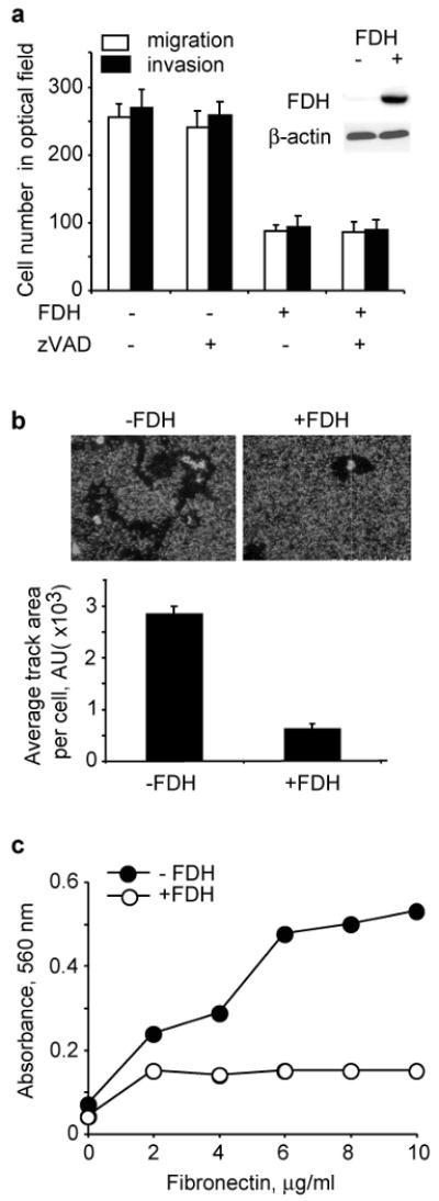
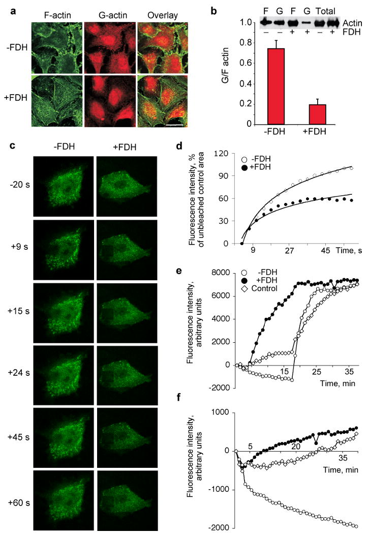
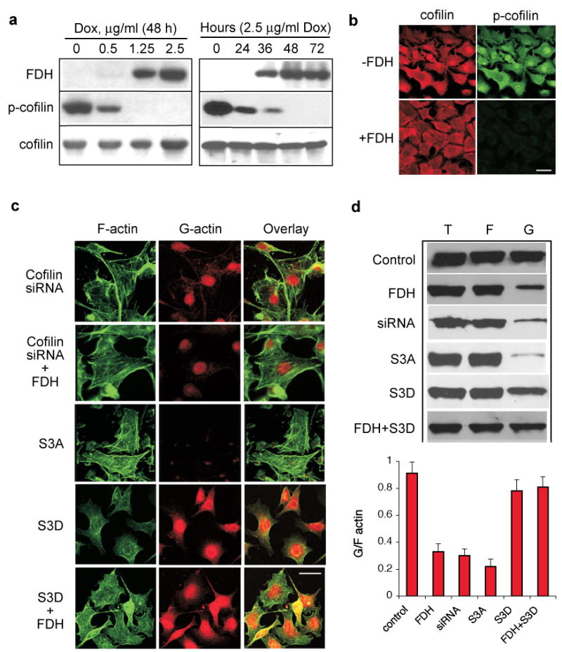
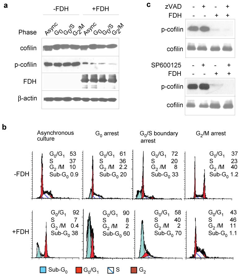
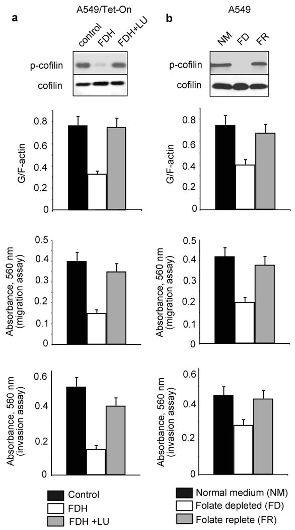
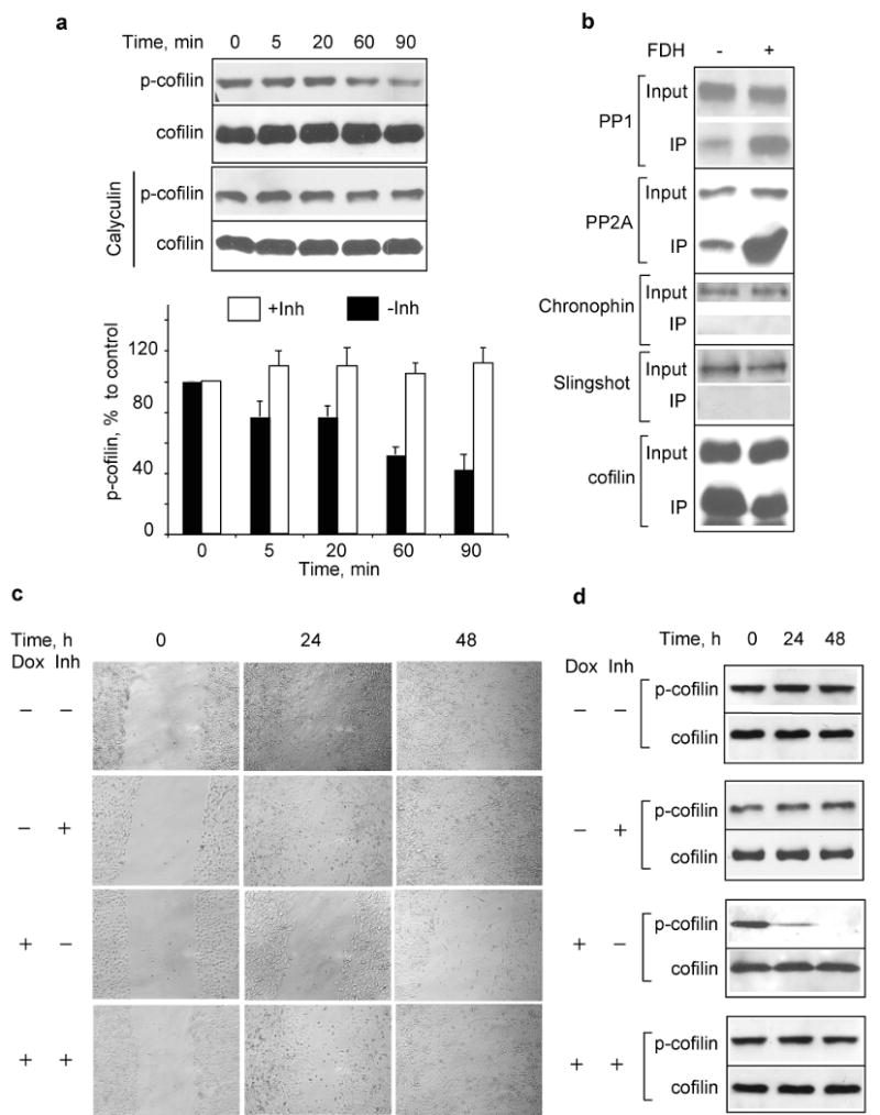
Here we report that ALDH1L1 (FDH, a folate enzyme with tumor suppressor-like properties) inhibits cell motility. The underlying mechanism involves F-actin stabilization, re-distribution of cytoplasmic actin toward strong preponderance of filamentous actin and formation of actin stress fibers. A549 cells expressing FDH showed a much slower recovery of green fluorescent protein-actin fluorescence in a fluorescence recovery after photobleaching assay, as well as an increase in G-actin polymerization and a decrease in F-actin depolymerization rates in pyren-actin fluorescence assays indicating the inhibition of actin dynamics. These effects were associated with robust dephosphorylation of the actin depolymerizing factor cofilin by PP1 and PP2A serine/threonine protein phosphatases, but not the cofilin-specific phosphatases slingshot and chronophin. In fact, the PP1/PP2A inhibitor calyculin prevented cofilin dephosphorylation and restored motility. Inhibition of FDH-induced apoptosis by the Jun N-terminal kinase inhibitor SP600125 or the pan-caspase inhibitor zVAD-fmk did not restore motility or levels of phosphor-cofilin, indicating that the observed effects are independent of FDH function in apoptosis. Interestingly, cofilin small interfering RNA or expression of phosphorylation-deficient S3A cofilin mutant resulted in a decrease of G-actin and the actin stress fiber formation, the effects seen upon FDH expression. In contrast, the expression of S3D mutant, mimicking constitutive phosphorylation, prevented these effects further supporting the cofilin-dependent mechanism. Dephosphorylation of cofilin and inhibition of motility in response to FDH can also be prevented by the increased folate in media. Furthermore, folate depletion itself, in the absence of FDH, resulted in cofilin dephosphorylation and inhibition of motility in several cell lines. Our experiments showed that these effects were folate specific and not a general response to nutrient starvation. Overall, this study shows the presence of distinct intracellular signaling pathways regulating motility in response to folate status and points toward mechanisms involving folates in promoting a malignant phenotype.
Conflict of interest statement
Figures
References
-
- Ambach A, Saunus J, Konstandin M, Wesselborg S, Meuer SC, Samstag Y. The serine phosphatases PP1 and PP2A associate with and activate the actin-binding protein cofilin in human T lymphocytes. Eur J Immunol. 2000;30:3422–31. - PubMed
-
- Ballestrem C, Wehrle-Haller B, Imhof BA. Actin dynamics in living mammalian cells. J Cell Sci. 1998;111(Pt 12):1649–58. - PubMed
-
- Bamburg JR, Wiggan OP. ADF/cofilin and actin dynamics in disease. Trends Cell Biol. 2002;12:598–605. - PubMed
-
- Chanson A, Sayd T, Rock E, Chambon C, Sante-Lhoutellier V, Potier de Courcy G, et al. Proteomic analysis reveals changes in the liver protein pattern of rats exposed to dietary folate deficiency. J Nutr. 2005;135:2524–9. - PubMed
Publication types
MeSH terms
Substances
Grants and funding
LinkOut - more resources
Full Text Sources
Research Materials
Miscellaneous
