Review
. 2008;31(4):310-39.

How adaptation of the brain to alcohol leads to dependence: a pharmacological perspective

Review

How adaptation of the brain to alcohol leads to dependence: a pharmacological perspective

Peter Clapp et al. Alcohol Res Health. 2008.

Abstract

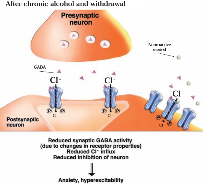
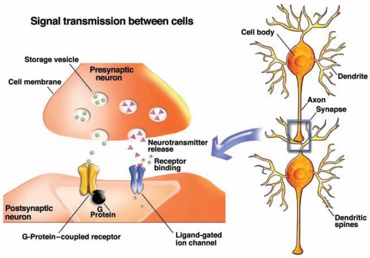
The development of alcohol dependence is posited to involve numerous changes in brain chemistry (i.e., neurotransmission) that lead to physiological signs of withdrawal upon abstinence from alcohol as well as promote vulnerability to relapse in dependent people. These neuroadaptive changes often occur in those brain neurotransmission systems that are most sensitive to the acute, initial effects of alcohol and/or contribute to a person’s initial alcohol consumption. Studies of these neuroadaptive changes have been aided by the development of animal models of alcohol dependence, withdrawal, and relapse behavior. These animal models, as well as findings obtained in humans, have shed light on the effects that acute and chronic alcohol exposure have on signaling systems involving the neurotransmitters glutamate, γaminobutyric acid (GABA), dopamine, and serotonin, as well as on other signaling molecules, including endogenous opioids and corticotrophin-releasing factor (CRF). Adaptation to chronic alcohol exposure by these systems has been associated with behavioral effects, such as changes in reinforcement, enhanced anxiety, and increased sensitivity to stress, all of which may contribute to relapse to drinking in abstinent alcoholics. Moreover, some of these systems are targets of currently available therapeutic agents for alcohol dependence.


Figures

Figure 1
Figure 1
Location of some of the regions in the human brain that are affected by alcohol, including the mesolimbic dopamine system (which includes the ventral tegmental area [VTA], nucleus accumbens, and prefrontal cortex), amygdala, striatum, and hippocampus.
Figure 2A
Figure 2A
Actions of the brain’s glutamate system. Glutamate (green circles) exerts its effects by acting on various types of receptors, including the N-methyl-d-aspartate receptors (NMDARs) and α-amino-3-hydroxy-5-methylisoxazole-4-proprionic acid receptors (AMPARs), both of which are ion channels, and metabotropic glutamate receptors (mGluRs), which are coupled to G-proteins. G-proteins, in turn, indirectly activate protein kinase C (PKC) and activate or inhibit adenyl cyclase (AC), depending on the mGluR and G-protein involved. In the absence of alcohol, glutamate leads to the activation of the postsynaptic neuron and the generation of a new nerve signal.
Figure 2B
Figure 2B
Actions of the brain’s glutamate system. In the presence of alcohol (ethanol, purple circles), the activity of the N-methyl-d-aspartate receptors (NMDARs) and α-amino-3-hydroxy-5-methylisoxazole-4-proprionic acid receptors (AMPARs), is inhibited, reducing cation entry into the cell. As a result, the activity of the neuron is reduced and no or fewer nerve signals are generated. For further information, see legend to figure 2A.
Figure 2C
Figure 2C
Actions of the brain’s glutamate system. After chronic alcohol exposure and during withdrawal, glutamate release at the synapse is enhanced and the number of synaptic N-methyl-d-aspartate receptors (NMDARs) and α-amino-3-hydroxy-5-methylisoxazole-4-proprionic acid receptors (AMPARs) is increased. As a result, glutamate induces excessive activity of the postsynaptic neuron. For further information, see legend to figure 2A.
Figure 3
Figure 3
Lengthwise view of the rat brain showing the distribution of opioid peptide–producing neurons. The opioid peptides—endorphins (teal), enkephalins (purple), and dynorphins (blue)—and the neurotransmitter dopamine are involved in the processes of reward and reinforcement. Endorphin-producing neurons are located primarily in the arcuate nucleus (ArcN) of the hypothalamus and the nucleus tractus solitarius (NTS); they extend to and release endorphin in various brain areas (purple). Nerve cells in several regions produce enkephalins and dynorphins, which may be released either in the same region or in distant regions through networks of neurons (not shown). The mesolimbic dopamine system (orange line) is influenced by the actions of endogenous opioids and carries dopamine from the ventral tegmental area (VTA) to various parts of the brain (see also figure 1). NOTE: Amyg = amygdala; CPu = caudate putamen; FC = frontal cortex; Hpc = hippocampus; NAc = nucleus accumbens; PaG = periaqueductal gray area; Sept = septum. SOURCE: Gianoulakis, C. Alcohol-seeking behavior: The roles of the hypothalamic-pituitary-adrenal axis and the endogenous opioid system. Alcohol Health & Research World 22(3):202–210, 1998. PMID: 15706797
Figure 4
Figure 4
Alcohol’s effects on endogenous opioids and the mesolimbic dopamine system. The activity of the dopamine-releasing (i.e., dopaminergic) neurons in the ventral tegmental area (VTA) is controlled by γ –aminobutyric acid (GABA)-releasing (i.e., GABAergic) neurons. When these GABA neurons are activated (e.g., through the actions of the excitatory neuro-transmitter glutamate), their signals decrease the firing of dopaminergic neurons. Endogenous opioids, however, can act on μ receptors on the GABAergic neurons, thereby inhibiting GABA transmission, and ultimately leading to increased dopamine release. A) Acute alcohol can induce β-endorphin release, resulting in activation of μ receptors on the GABAergic neurons in VTA. This, in combination with alcohol’s inhibition of glutamate effects on GABA neurons, could lead to decreased GABAergic activity in the VTA, and subsequently increased firing of the dopaminergic neurons, resulting in increased dopamine release in the nucleus accumbens (NAc). Alcohol also directly increases the activity of dopamine neurons. B) During withdrawal from alcohol, after chronic alcohol exposure that produces alcohol dependence (i.e., in the absence of alcohol in a dependent individual), glutamate input to GABA neurons is increased, leading to decreased dopamine release. In addition, the activity of the VTA dopamine neurons is reduced. C) When alcohol is reintroduced, the dopamine neurons are more sensitive to alcohol’s direct effects; moreover, alcohol again inhibits glutamate β-endor-phin release, thereby reversing the decreased dopamine release that occurs in the alcohol-abstinent, alcohol-dependent individual. NOTE: Other systems that interact with alcohol to control dopamine neuron activity in the VTA (and dopamine release in the nucleus accumbens), but that are not shown in this figure, include endogenous cannabinoids (which can affect GABA release and interact with opioid systems), nicotinic cholinergic receptors, and serotonin transmission.
Figure 5A
Figure 5A
Actions of the brain’s γ-aminobutyric acid (GABA) system. GABA acts in part through GABAA receptors, which serve as ion channels for chloride ions (Cl). Greater influx of Cl into the neuron makes it more difficult for the cell to generate a new nerve impulse.
Figure 5B
Figure 5B
Actions of the brain’s γ-aminobutyric acid (GABA) system. In the presence of ethanol, GABA activity is enhanced, resulting in greater Cl influx into the postsynaptic neuron and, consequently, greater inhibition of the neuron. (For more information, see legend to figure 5A.)
Figure 5C
Figure 5C
Actions of the brain’s γ-aminobutyric acid (GABA) system. After chronic alcohol exposure and during withdrawal, GABA activity at the synapse is reduced, leading to reduced inhibition of the postsynaptic neuron. This results in development of anxiety and hyperexcitability. (For more information, see legend to figure 5A.)
None

References

    1. Ait-Daoud N, Johnson BA, Javors M, et al. Combining ondansetron and naltrexone treats biological alcoholics: Corroboration of self-reported drinking by serum carbohydrate deficient transferrin, a biomarker. Alcoholism: Clinical and Experimental Research. 2001;25:847–849. - PubMed
    1. Boyce-Rustay JM, Wiedholz LM, Millstein RA, et al. Ethanol-related behaviors in serotonin transporter knockout mice. Alcoholism: Clinical and Experimental Research. 2006;30:1957–1965. - PubMed
    1. Casu MA, Pisu C, Lobina C, Pani L. Immunocytochemical study of the forebrain serotonergic innervation in Sardinian alcohol-preferring rats. Psychopharmacology (Berlin) 2004;172:341–351. - PubMed
    1. Colombo G, Orru A, Lai P, et al. The cannabinoid CB1 receptor antagonist, rimonabant, as a promising pharmacotherapy for alcohol dependence: Preclinical evidence. Molecular Neurobiology. 2007;36:102–112. - PubMed
    1. Hodge CW, Kelley SP, Bratt AM, et al. 5-HT(3A) receptor subunit is required for 5-HT3 antagonist-induced reductions in alcohol drinking. Neuropsychopharmacology. 2004;29:1807–1813. - PubMed

Publication types

MeSH terms

LinkOut - more resources