

Using noncontrast cardiac CT and coronary artery calcification measurements for cardiovascular risk assessment and management in asymptomatic adults
- PMID: 20730074
- PMCID: PMC2922319
- DOI: 10.2147/vhrm.s7457
Using noncontrast cardiac CT and coronary artery calcification measurements for cardiovascular risk assessment and management in asymptomatic adults
Abstract
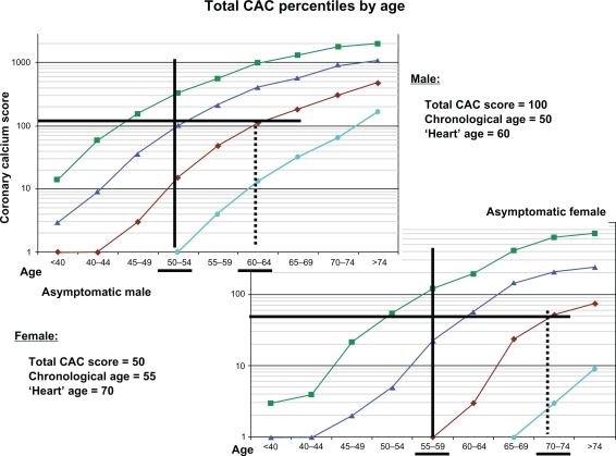
The presence of mural calcification has, for decades, been recognized as a marker for atheromatous plaque in the coronary arteries and the aorta, but only in the past decade has the application of noncontrast computed tomography (CT) been shown to be a reproducible, safe, and convenient test, which now is available worldwide. However, awareness of coronary artery calcium scanning is insufficient and the practitioner must be aware of the available literature as well as understanding clinical recommendations for applications and interpretation. It is best applied in the medium/intermediate risk, asymptomatic adult regardless of ethnicity across broad age ranges for both men and women; additional prognostic information is also afforded from the calcium distribution in the coronary artery system. Additionally, information can also be derived from the same CT scan regarding heart and aorta size and assessment of the epicardial fat pad (an anatomic marker for the metabolic syndrome). Details of how this test can aid in cardiovascular risk assessment and management in adults are provided.
Keywords: National Cholesterol Education Program Adult Treatment Plan III (NCEP ATP III); coronary artery calcium; coronary artery disease; electron beam computed tomography; epicardial fat; multidetector computed tomography.
Figures

Similar articles
-
Spiral computed tomography evidence of close correlation between coronary and thoracic aorta calcifications.Atherosclerosis. 2004 Sep;176(1):133-8. doi: 10.1016/j.atherosclerosis.2004.03.027. Atherosclerosis. 2004. PMID: 15306185
-
Progression of coronary artery calcification and thoracic aorta calcification in kidney transplant recipients.Am J Kidney Dis. 2012 Feb;59(2):258-69. doi: 10.1053/j.ajkd.2011.07.019. Epub 2011 Sep 23. Am J Kidney Dis. 2012. PMID: 21944666
-
Thoracic aortic calcium versus coronary artery calcium for the prediction of coronary heart disease and cardiovascular disease events.JACC Cardiovasc Imaging. 2009 Mar;2(3):319-26. doi: 10.1016/j.jcmg.2008.12.010. JACC Cardiovasc Imaging. 2009. PMID: 19356578
-
Electron beam computed tomographic coronary calcium scanning: a review and guidelines for use in asymptomatic persons.Mayo Clin Proc. 1999 Mar;74(3):243-52. doi: 10.4065/74.3.243. Mayo Clin Proc. 1999. PMID: 10089993 Review.
-
Expert review on coronary calcium.Vasc Health Risk Manag. 2008;4(2):315-24. doi: 10.2147/vhrm.s1160. Vasc Health Risk Manag. 2008. PMID: 18561507 Free PMC article. Review.
Cited by
-
The Relationship between Job Strain and Ischemic Heart Disease Mediated by Endothelial Dysfunction Markers and Imaging.Medicina (Kaunas). 2024 Jun 26;60(7):1048. doi: 10.3390/medicina60071048. Medicina (Kaunas). 2024. PMID: 39064476 Free PMC article.
-
Ex vivo coronary calcium volume quantification using a high-spatial-resolution clinical photon-counting-detector computed tomography.J Med Imaging (Bellingham). 2023 Jul;10(4):043501. doi: 10.1117/1.JMI.10.4.043501. Epub 2023 Jul 4. J Med Imaging (Bellingham). 2023. PMID: 37408984 Free PMC article.
-
Serum Homocysteine and Vascular Calcification: Advances in Mechanisms, Related Diseases, and Nutrition.Korean J Fam Med. 2022 Sep;43(5):277-289. doi: 10.4082/kjfm.21.0227. Epub 2022 Sep 20. Korean J Fam Med. 2022. PMID: 36168899 Free PMC article.
-
The correlation of epicardial adipose tissue on postmortem CT with coronary artery stenosis as determined by autopsy.Forensic Sci Med Pathol. 2015 Jun;11(2):186-92. doi: 10.1007/s12024-015-9659-7. Epub 2015 Feb 25. Forensic Sci Med Pathol. 2015. PMID: 25711291
-
Reliable categorisation of visual scoring of coronary artery calcification on low-dose CT for lung cancer screening: validation with the standard Agatston score.Eur Radiol. 2013 May;23(5):1226-33. doi: 10.1007/s00330-012-2726-5. Epub 2012 Dec 14. Eur Radiol. 2013. PMID: 23239060
References
-
- Heart and Stroke Statistical Update. Dallas, TX: American Heart Association; 2001.
-
- Falk E, Shah PK, Fuster V. Coronary plaque disruption. Circulation. 1995;92:657–671. - PubMed
-
- Virchow R. Die Cellulärpathologie in Ihrer Begrundung auf physiologische und pathologische Gewebesslehre. Berlin: August Hirschwald; 1858.
-
- Blankenhorn DH. Coronary arterial calcification: a review. Am J Med Sci. 1961;242:1–9.
-
- Tannenbaum SR, Kondos GT, Veselik KE, Prendergast MR, Brundage BH, Chomka EV. Detection of calcific deposits in coronary arteries by ultrafast computed tomography and correlation with angiography. Am J Cardiol. 1989;63:870–872. - PubMed
Publication types
MeSH terms
LinkOut - more resources
Full Text Sources
Medical
Miscellaneous
