. 2010 Aug 13;5(8):e12173.
doi: 10.1371/journal.pone.0012173.

Conserved and distinct modes of CREB/ATF transcription factor regulation by PP2A/B56gamma and genotoxic stress

Affiliations

Conserved and distinct modes of CREB/ATF transcription factor regulation by PP2A/B56gamma and genotoxic stress

Naval P Shanware et al. PLoS One. .

Abstract

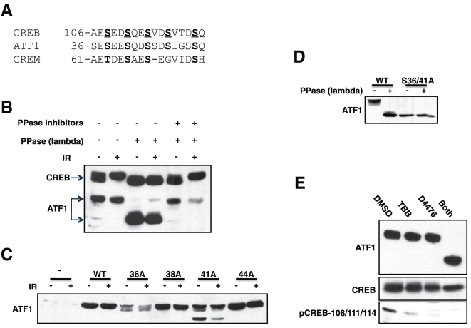
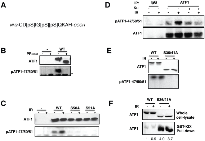
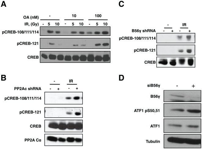
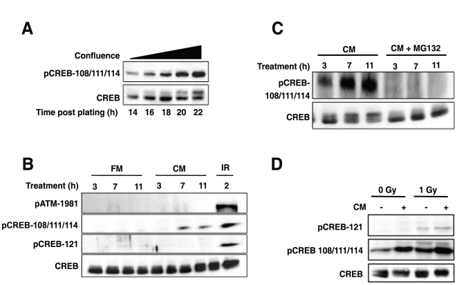
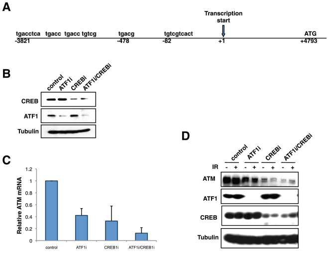
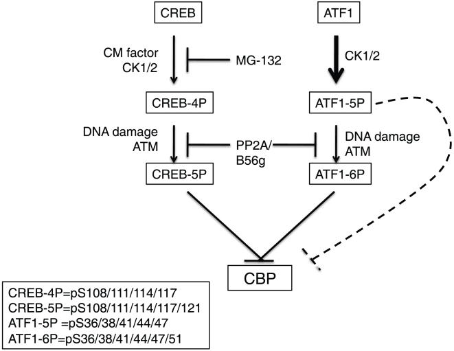
Activating transcription factor 1 (ATF1) and the closely related proteins CREB (cyclic AMP resonse element binding protein) and CREM (cyclic AMP response element modulator) constitute a subfamily of bZIP transcription factors that play critical roles in the regulation of cellular growth, metabolism, and survival. Previous studies demonstrated that CREB is phosphorylated on a cluster of conserved Ser residues, including Ser-111 and Ser-121, in response to DNA damage through the coordinated actions of the ataxia-telangiectasia-mutated (ATM) protein kinase and casein kinases 1 and 2 (CK1/2). Here, we show that DNA damage-induced phosphorylation by ATM is a general feature of CREB and ATF1. ATF1 harbors a conserved ATM/CK cluster that is constitutively and stoichiometrically phosphorylated by CK1 and CK2 in asynchronously growing cells. Exposure to DNA damage further induced ATF1 phosphorylation on Ser-51 by ATM in a manner that required prior phosphorylation of the upstream CK residues. Hyperphosphorylated ATF1 showed a 4-fold reduced affinity for CREB-binding protein. We further show that PP2A, in conjunction with its targeting subunit B56gamma, antagonized ATM and CK1/2-dependent phosphorylation of CREB and ATF1 in cellulo. Finally, we show that CK sites in CREB are phosphorylated during cellular growth and that phosphorylation of these residues reduces the threshold of DNA damage required for ATM-dependent phosphorylation of the inhibitory Ser-121 residue. These studies define overlapping and distinct modes of CREB and ATF1 regulation by phosphorylation that may ensure concerted changes in gene expression mediated by these factors.


Conflict of interest statement

Competing Interests: The authors have declared that no competing interests exist.

Figures

Figure 1
Figure 1. ATF1 is constitutively phosphorylated by CK1/CK2 in vivo.
(A) Sequence overlay of ATM/CK cluster regions in CREB, CREM and ATF1. Homologous putative phosphorylation sites are shown in boldface and defined phosphorylation sites in CREB underlined. (B) ATF1 is basally phosphorylated in intact cells. HEK 293T cells were exposed to IR (10 Gy) or left untreated and cell extracts were prepared and treated with λ phosphatase (with or without inhibitors) prior to analysis by SDS-PAGE followed by immunoblotting with α-bZIP antibody that recognizes CREB and ATF1. The * denotes the position of a cross-reactive protein. (C) Phosphorylation site requirements for ATF1 electrophoretic mobility shift. HEK 293T cells were transfected with plasmids encoding Myc-ATF1WT or the indicated Myc-ATF1 phosphorylation site mutants. Cell extracts were then made and analyzed by immunoblotting using α-Myc antibody. (D) The ATF1S36/41A mimics in vitro dephosphorylated ATF1. HEK 293T cells were transfected with plasmids encoding Myc-ATF1WT or the Myc-ATF1S36/41A mutant. Cell extracts were prepared and treated with λ phosphatase prior to analysis by immunoblotting using α-Myc antibodies. (E) CK1 and CK2 inhibitors dephosphorylate ATF1. HEK 293T cells were treated with 75 µM D4476, 50 µM TBB or both compounds for 4 h. Cell extracts were then analyzed by immunoblotting using α-ATF1, α-CREB and α-pCREB-108/111/114 antibodies.
Figure 2
Figure 2. DNA damage induces ATM dependent phosphorylation of ATF1 on Ser-51.
(A) Sequence of peptide antigen used to generate α-pATF1-47/50/51 antibody. (B) Phosphatase sensitivity of α-pATF1-47/50/51 antibody. HEK 293T cells were transfected with Myc-ATF1WT plasmid. Cell extracts were prepared and treated with λ phosphatase prior to analysis by immunoblotting using α-Myc and α-pATF1-47/50/51 antibodies. (C) Phosphorylation site requirements and IR inducibility. HEK 293T cells were transfected with vector DNA (−), Myc-ATF1WT, Myc-ATF1S50A or Myc-ATF1S51A plasmids and exposed to IR (10 Gy, 2 h). Cell extracts were prepared and analyzed by immunoblotting using α-Myc and α-pATF1-47/50/51 antibodies. (D) IR dependent ATF1 phosphorylation is ATM dependent. HEK 293T cells either left untreated or treated with IR in the presence of 10 µM ATM inhibitor (KU-55933). Immunoprecipitation reactions were performed using a mock antibody or α-ATF1 antibody and immunoprecipitates were analyzed by immunoblotting using α-Myc and α-pATF1-47/50/51 antibodies. (E) The ATF1S36/41A mutant is defective for IR induced pATF1-47/50/51 phosphorylation. HEK 293T cells were transfected with plasmid DNA encoding Myc-ATF1WT or the Myc-ATF1S36/41A mutant and either left untreated or subjected to 10 Gy IR for 2 h. Cell extracts were prepared and analyzed by immunoblotting using α-Myc and α-pATF1-47/50/51 antibodies. (F) Hyperphosphorylated ATF1 shows reduced binding to the KIX domain of CBP. HEK 293T cells were transfected with plasmids encoding Myc-ATF1WT or the Myc-ATF1S36/41A mutant and either left untreated or subjected to 10 Gy IR. Cell extracts were prepared 2 h later, incubated with GST-KIX-loaded beads, and bound protein analyzed by immunoblotting using α-Myc antibodies. Numbers under GST-KIX result demote fold changes in ATF1 levels.
Figure 3
Figure 3. B56γ-PP2A mediates dephosphorylation of CREB and ATF1.
(A) Okadaic acid (OA) sensitivity. HEK 293T cells were left untreated or exposed to 10 nM and 100 nM OA for 1 h. They were then subjected to IR for the indicated times. Cell extracts were prepared and analyzed by immunoblotting with α-CREB, α-pCREB-108/111/114 and α-pCREB-121 antibodies. (B) PP2Ac knockdown stimulates DNA damage-dependent CREB phosphorylation. HEK 293T cells expressing an shRNA targeting PP2Ac were compared to cells expressing a non-targeting shRNA construct. Cells were exposed to 10 Gy IR for 2 h and subjected to immunoblotting analysis with α-CREB, α-pCREB-108/111/114, α-pCREB-121 and α-PP2Ac antibodies. (C) B56γ knockdown stimulates DNA damage-dependent CREB phosphorylation. HEK 293T cells expressing an shRNA targeting B56γ were compared to cells expressing a non-targeting shRNA construct. Cells were exposed to 10 Gy IR for 1 h and subjected to immunoblotting analysis with α-CREB, α-pCREB-108/111/114, α-pCREB-121 and α-B56γ antibodies. (D) Effects of B56γ knockdown on ATF1 Ser-47/50/51 phosphorylation. HEK 293T cells were transiently transfected with control or B56γ siRNA and the levels of ATF1 Ser-47/50/51 phosphorylation assessed using α-pATF1-47/50/51 antibodies.
Figure 4
Figure 4. ATM and DNA damage-independent phosphorylation of the CREB ATM/CK cluster during cell growth.
(A) CREB phosphorylation increases with time spent in culture. Replicate plates of HEK 293T cells were plated at 60–70% confluence and allowed to grow for 14 h. Cells were then harvested at 2 h intervals and the extracts analyzed by immunoblotting with α-CREB and α-pCREB-108/111/114 antibodies. (B) Conditioned media (CM) induces CREB phosphorylation independent of DNA damage. HEK 293T cells were plated overnight followed by exposure to fresh media (FM), or CM. A 10 Gy IR exposure (2 h) was used as a positive control to induce Ser-121 phosphorylation. Cell extracts were prepared at the indicated times and analyzed by immunoblotting with α-CREB, α-pCREB-108/111/114, α-pCREB-121, α-ATM and α- pATM-1981 antibodies. (C) MG-132 suppresses CM-induced CREB phosphorylation. HEK 293T cells were cultured in the presence of CM or CM supplemented with 10 µM MG132 (or solvent). Cell extracts were prepared at the indicated times after CM treatment and analyzed by immunoblotting with α-CREB and α-pCREB-108/111/114 antibodies. (D) CM-induced phosphorylation of CREB on Ser-111 facilitates IR-induced phosphorylation of Ser-121. HEK 293T cells were incubated with CM or not for 8 h and then mock irradiated or exposed to 1 Gy of IR. Cell extracts were analyzed by immunoblotting with α-CREB, α-pCREB-108/111/114 and α-pCREB-121 antibodies.
Figure 5
Figure 5. CREB/ATF1 regulate ATM transcription.
(A) Schematic of the human ATM with putative CREs is shown. CREs conserved between human and mouse ATM promoters are in boldface. The arrow indicates the transcription start site. (B) Knockdown of CREB and ATF1 in MeWo cells. MeWo cells were transfected with shRNA for ATF1, siRNA for CREB or both and compared to cells expressing a non-targeting construct for 48 h. Cell extracts were subjected to immunoblotting with α-CREB, α-ATF1 and α-β-Tubulin antibodies. (C) Real-time PCR analysis of ATM mRNA in CREB/ATF1-deficient MeWo cells. Relative fold-expression levels of ATM mRNA normalized for GAPDH is shown. Error bars denote standard deviation from the mean from three independent experiments. (D) Effects of CREB and ATF1 knockdown on ATM protein levels. MeWo cells were transfected with shRNA for ATF1, siRNA for CREB or both and compared to cells expressing a non-targeting construct for 48 h before IR exposure. Cell extracts were prepared and subjected to immunoblotting with α-CREB, α-ATF1, α-ATM and α-β-Tubulin antibodies.
Figure 6
Figure 6. Working model depicting regulation of CREB and ATF1 on ATM/CK cluster residues.
Both proteins are constitutively phosphorylated on CK residues resulting in CREB-4P and ATF1-5P isoforms that are further phosphorylated in response to DNA damage on Ser-121 and Ser-51, respectively to yield CREB-5P and ATF1-6P isoforms. Phosphorylation of CREB CK residues (Ser-108/111/114/117) is stimulated by a CM factor, whereas ATF1 CK residues (Ser-36/38/41/44/47) are constitutively phosphorylated (indicated by bold arrow). PP2A/B56γ antagonizes phosphorylation of ATM sites in both CREB and ATF1. Inhibition of CBP binding is one endpoint of ATM/CK cluster phosphorylation. The dashed line denotes that phosphorylation of CK sites in ATF1 is sufficient to inhibit CBP binding in the absence of DNA damage.

Similar articles

Cited by

References

    1. Mayr B, Montminy M. Transcriptional regulation by the phosphorylation-dependent factor CREB. Nat Rev Mol Cell Biol. 2001;2:599–609. - PubMed
    1. Zhang X, Odom DT, Koo SH, Conkright MD, Canettieri G, et al. Genome-wide analysis of cAMP-response element binding protein occupancy, phosphorylation, and target gene activation in human tissues. Proc Natl Acad Sci U S A. 2005;102:4459–4464. - PMC - PubMed
    1. Impey S, McCorkle SR, Cha-Molstad H, Dwyer JM, Yochum GS, et al. Defining the CREB regulon: a genome-wide analysis of transcription factor regulatory regions. Cell. 2004;119:1041–1054. - PubMed
    1. Montminy M, Koo SH, Zhang X. The CREB family: key regulators of hepatic metabolism. Ann Endocrinol (Paris) 2004;65:73–75. - PubMed
    1. Lonze BE, Ginty DD. Function and regulation of CREB family transcription factors in the nervous system. Neuron. 2002;35:605–623. - PubMed

Publication types

MeSH terms