. 2011 Mar;68(6):1041-51.
doi: 10.1007/s00018-010-0503-7. Epub 2010 Aug 22.

Cytoprotective effects of acidosis via heat shock protein HSP27 against the anticancer drug doxorubicin

Affiliations

Cytoprotective effects of acidosis via heat shock protein HSP27 against the anticancer drug doxorubicin

Anurag Kumar Singh et al. Cell Mol Life Sci. 2011 Mar.

Abstract

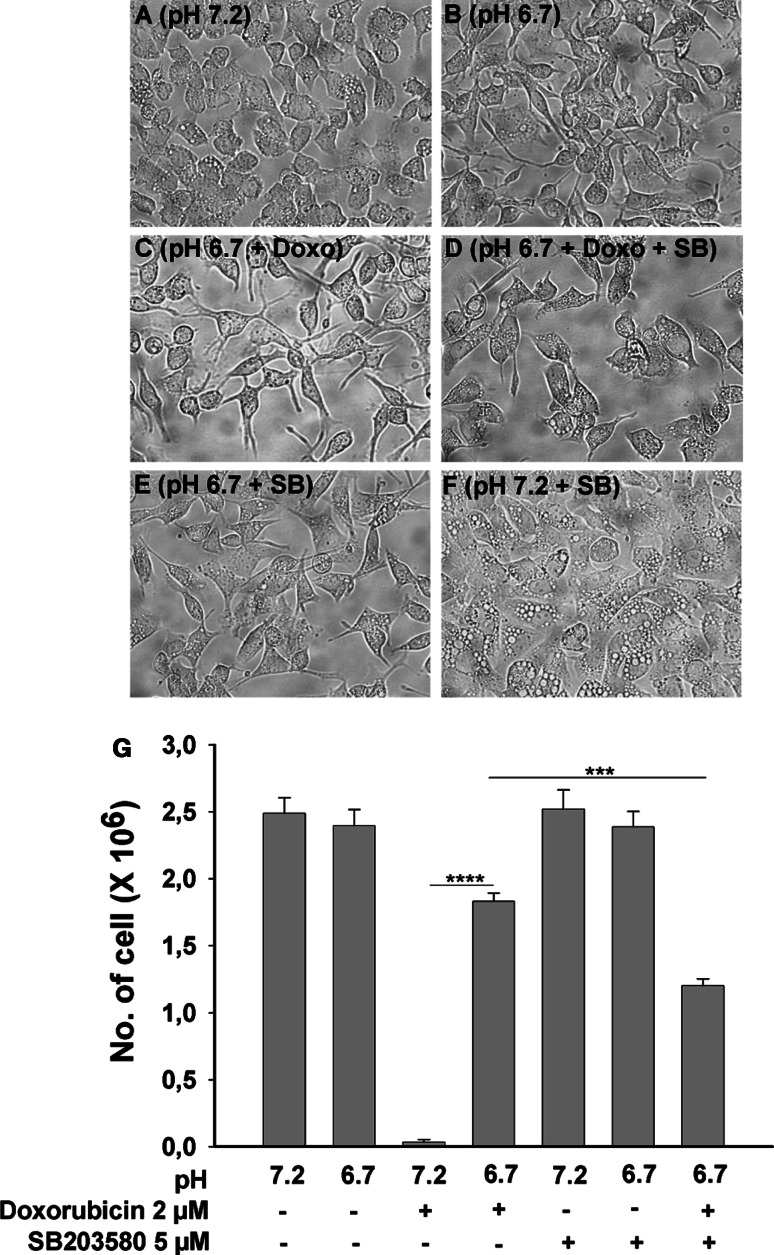
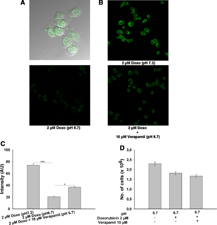
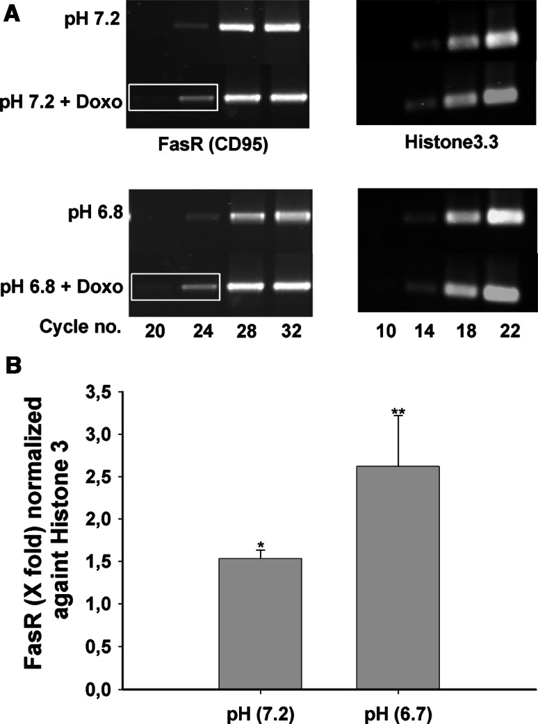
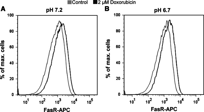
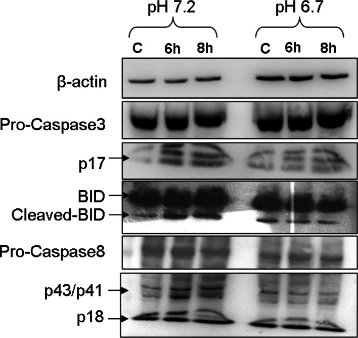
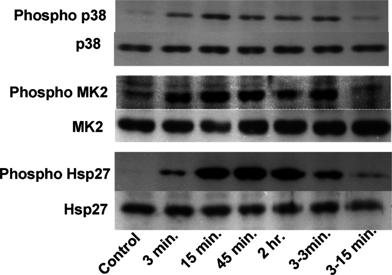
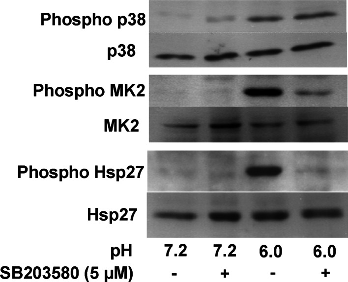
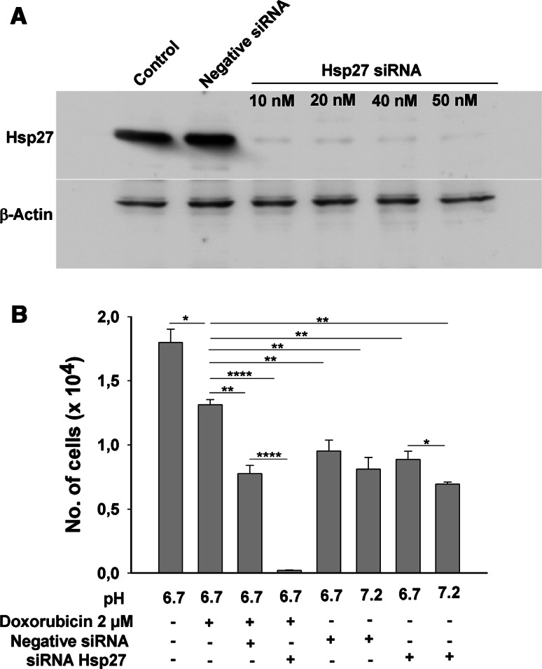
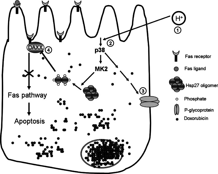
Drug resistance continues to be a stumbling block in achieving better cure rates in several cancers. Doxorubicin is commonly used in treatment of a wide range of cancers. The aim of this study was to look into the mechanisms of how low ambient pH may contribute to down-regulation of apoptotic pathways in a gastric tumour cell line. Low pH culture conditions were found to dramatically prolong cell survival after doxorubicin treatment, an effect that was in part reversed by co-incubation with the specific p38 mitoge-activated protein kinase (MAP kinase) inhibitor SB203580, only mildly inhibited by blockade of the multi-drug resistance 1 (MDR1) transporter, but completely abolished by siRNA-mediated knockdown of the heat shock protein 27 (HSP27). In conclusion, acidic pH causes less accumulation of cytotoxic drug in the nucleus of adeno gastric carcinoma (AGS) cells and HSP27-dependent decrease in FasR-mediated gastric epithelial tumour cell apoptosis.


Figures

Fig. 1
Fig. 1
Low ambient pH rescues AGS cells from doxorubicin-induced cell death. Morphology of the cells cultured under various conditions. 2 × 106 AGS cell were cultured in serum free condition for 48 h. Cells cultured at physiological pH (pH 7.2) showed some vacuolization (a), which was not present at pH 6.7 (b), seen much more pronounced in the presence of SB203580 (f), and was largely prevented by concomitant incubation at pH 6.7 (e). In the presence of doxorubicin, cells did not survive at pH 7.2 (g), whereas a large number of cells remained viable after 48 h in 2 μM doxorubicin at pH 6.7 (c,g). Co-incubation with SB203580 along with doxorubicin induced significantly higher number of cell death as compared to doxorubicin alone at pH 6.7 (d, g). g Bars indicate the total number of cells left after 48 h of culture. Results are ± SEM. *Significant low no. of cells between the two groups connected via horizontal line: ***<0.0001 and ****<0.00001
Fig. 2
Fig. 2
Intracellular doxorubicin concentration in cells incubated at physiological and low ambient pH. Overlay of transmission picture of cells and nuclei shows that most of the doxorubicin (green) is retained in the nucleus (a). The doxorubicin retention was significantly higher at pH 7.2 than at low pH, and co-incubation with 10 μM verapamil significantly increased the doxorubicin retention at lower pH (b). c Mean intensity from 35 nuclei in the various groups. d Despite significantly higher intracellular doxorubicin concentration, upon co-incubation with 10 μM verapamil along with 2 μM doxorubicin did not affect cell survival. Results are ± SEM. *Significant high amount of doxorubicin in the nuclei of cells connected via horizontal line: *<0.01 and ***<0.001
Fig. 3
Fig. 3
Up-regulation of FasR expression by doxorubicin in AGS cells. a The left panel shows the semiquantitative PCR for the FasR, with distinctly stronger bands at cycle no. 20 and 24 in the presence compared to the absence of 2 μM doxorubicin. The same up-regulation was seen when the cells were incubated at pH 6.7. Right panel shows the Histone3 band. b Densitometry calculation from cycle 24 of FasR and cycle 18 from the Histone3 show 1.5- and 2.6-fold difference at pH 7.2 and pH 6.7, respectively. Results are ± SEM. *Significantly higher value as compared to the control cells which did not get doxorubicin treatment: *≤0.01 and **≤0.001
Fig. 4
Fig. 4
Up-regulation of FasR expression by doxorubicin in AGS cells. a The left panel shows the histogram of FasR-APC of the cells processed at the pH 7.2. The cells which were treated with the doxorubicin for 3 h (black curve) show clearly more cells which were APC positive as compared to the control cells (grey curve). b Similar results were obtained when cells were processed at the acidic pH (pH 6.7)
Fig. 5
Fig. 5
Doxorubicin induces activation of BID, Caspase-8 and -3. The uppermost row show the expression of β-actin as a loading control. The third row from the top clearly shows that there is more activated caspase-3 protein as compared to the control in pH 7.2 group. At the same time, this up-regulation in activated caspase-3 upon doxorubicin treatment was much greater as compared to the cells at the acidic pH (pH 6.7). The total caspase-3 level remains more or less the same (second row). The bottom two rows indicate the expression to total and activated (cleaved) caspase-8. The total protein remains more or less same and at the same time there was not that much difference in the activated caspase-8 (p18) expression. The other cleaved version of caspase-8 looks a little bit elevated at pH 7.2, while the expression of all the three bands looks very similar to pH 6.7. Expression of total BID protein remains more or less same in all the six lanes. The cleaved (activated) version shows a small amount of up-regulation only when incubated at pH 7.2
Fig. 6
Fig. 6
Incubation at low pH results in a rapid and sustained phosphorylation of p38 MAP kinase, the downstream target MK2, and HSP27. Time kinetics for AGS cells were exposed to acid (pH 6.0) for the indicated time periods and phospho-p38, phosphr MK2 and phosphoHSP27 levels was measured by western blot. Total p38, MK2 and HSP27 was taken as loading control, and demonstrated that acid exposure did not change the amount of these proteins. The time kinetics experiment shows that even in the presence of stimuli the level of phosphorylated kinase comes down after 45 min. A short exposure of stimuli for 3 min did not have a long-lasting effect (3–3 min and 3–15 min). Level of increased phosphorylated kinase was down to the level of control after 15 min when stimulus was taken off after 3 min
Fig. 7
Fig. 7
Acid-induced phosphorylation of HSP27 is p38 dependent. AGS cells were incubated at pH 7.2 and 6.0 for 10 min in the absence and presence of SB203580, a specific inhibitor of p38 MAP Kinase. The presence of SB203580 did not alter acid-induced phosphorylation of p38, but strongly reduced that of MK2 and HSP27
Fig. 8
Fig. 8
siRNA knockdown of HSP27 abolishes acid-conveyed resistance against doxorubicin-induced cell death. a A concentration of 50 nM siRNA against the HSP27 was able to knock down ~80% of the total HSP27, whereas “Allstars Negative” siRNA had no effect on cellular HSP27 content. β-Actin was taken as loading control which is the same in all the lanes. b Furthermore, in the cell survival experiments (similar to Fig. 1), the cells transfected with negative siRNA had better survival as compared to that of siRNA HSP27-transfected cells. Results are ± SEM. *Significant low no. of cells between the two groups connected via horizontal line.:*<0.01, **<0.001 and ****<0.00001
Fig. 9
Fig. 9
Working hypothesis of low pH induced resistance of AGS cells against doxorubicin toxicity. Intracellular acidosis activates p38 MAP kinase pathway here p38 which phosphorylates HSP27 via MK2. Phospho-HSP27 can block the apoptosis by preventing the interaction of DAXX to the FasR and may have other as yet unrecognized protective functions. In addition, doxirubicin accumulation in AGS cells is strongly reduced at low ambient pH, and this is in part mediated via P-glycoprotein (MDR1)

Similar articles

Cited by

References

    1. Semenza GL, Artemov D, Bedi A, Bhujwalla Z, Chiles K, Feldser D, Laughner E, Ravi R, Simons J, Taghavi P, Zhong H. ‘The metabolism of tumours’: 70 years later. Novartis Found Symp. 2001;240:251–260. doi: 10.1002/0470868716.ch17. - DOI - PubMed
    1. Gatenby RA, Gawlinski ET. A reaction-diffusion model of cancer invasion. Cancer Res. 1996;56:5745–5753. - PubMed
    1. Gatenby RA, Gawlinski ET. The glycolytic phenotype in carcinogenesis and tumor invasion: insights through mathematical models. Cancer Res. 2003;63:3847–3854. - PubMed
    1. Griffiths JR. Are cancer cells acidic? Br J Cancer. 1991;64:425–427. doi: 10.1038/bjc.1991.326. - DOI - PMC - PubMed
    1. Shrode LD, Tapper H, Grinstein S. Role of intracellular pH in proliferation, transformation, and apoptosis. J Bioenerg Biomembr. 1997;29:393–399. doi: 10.1023/A:1022407116339. - DOI - PubMed

Publication types

MeSH terms