

Immune cell location and function during post-natal mammary gland development
- PMID: 20730636
- PMCID: PMC4204476
- DOI: 10.1007/s10911-010-9188-7
Immune cell location and function during post-natal mammary gland development
Abstract
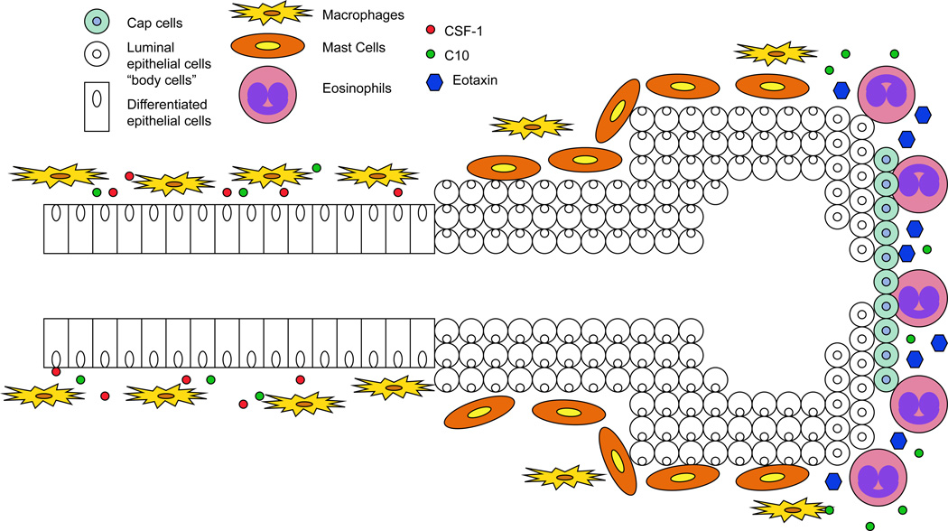
Post-natal mammary gland development requires complex interactions between the epithelial cells and various cell types within the stroma. Recent studies have illustrated the importance of immune cells and their mediators during the various stages of mammary gland development. However, the mechanisms by which these immune cells functionally contribute to mammary gland development are only beginning to be understood. This review provides an overview of the localization of immune cells within the mammary gland during the various stages of post-natal mammary gland development. Furthermore, recent studies are summarized that illustrate the mechanisms by which these cells are recruited to the mammary gland and their functional roles in mammary gland development.
Figures
References
-
- Watson CJ, Khaled WT. Mammary development in the embryo and adult: a journey of morphogenesis and commitment. Development. 2008;135(6):995–1003. - PubMed
-
- Regan MC, Kirk SJ, Wasserkrug HL, Barbul A. The wound environment as a regulator of fibroblast phenotype. J Surg Res. 1991;50(5):442–448. - PubMed
-
- Adamson R. Role of macrophages in normal wound healing: an overview. J Wound Care. 2009;18(8):349–351. - PubMed
-
- Barrientos S, Stojadinovic O, Golinko MS, Brem H, Tomic-Canic M. Growth factors and cytokines in wound healing. Wound Repair Regen. 2008;16(5):585–601. - PubMed
-
- Glaros T, Larsen M, Li L. Macrophages and fibroblasts during inflammation, tissue damage and organ injury. Front Biosci. 2009;14:3988–3993. - PubMed
Publication types
MeSH terms
Substances
Grants and funding
LinkOut - more resources
Full Text Sources
