

A device for separated and reversible co-culture of cardiomyocytes
- PMID: 20730771
- PMCID: PMC4031319
- DOI: 10.1002/btpr.431
A device for separated and reversible co-culture of cardiomyocytes
Abstract
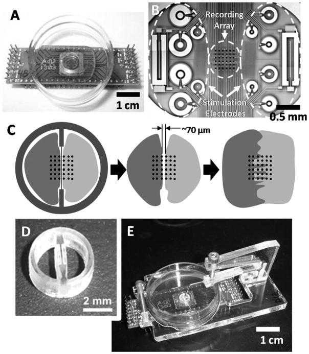
A novel technique is introduced for patterning and controllably merging two cultures of adherent cells on a microelectrode array (MEA) by separation with a removable physical barrier. The device was first demonstrated by separating two cardiomyocyte populations, which upon merging synchronized electrical activity. Next, two applications of this co-culture device are presented that demonstrate its flexibility as well as outline different metrics to analyze co-cultures. In a differential assay, the device contained two distinct cell cultures of neonatal wild-type and beta-adrenergic receptor (beta-AR) knockout cardiomyocytes and simultaneously exposed them with the beta-AR agonist isoproterenol. The beat rate and action potential amplitude from each cell type displayed different characteristic responses in both unmerged and merged states. This technique can be used to study the role of beta-receptor signaling and how the corresponding cellular response can be modulated by neighboring cells. In the second application, action potential propagation between modeled host and graft cell cultures was shown through the analysis of conduction velocity across the MEA. A co-culture of murine cardiomyocytes (host) and murine skeletal myoblasts (graft) demonstrated functional integration at the boundary, as shown by the progression of synchronous electrical activity propagating from the host into the graft cell populations. However, conduction velocity significantly decreased as the depolarization waves reached the graft region due to a mismatch of inherent cell properties that influence conduction.
(c) 2010 American Institute of Chemical Engineers
Figures


Similar articles
-
Modeling conduction in host-graft interactions between stem cell grafts and cardiomyocytes.Annu Int Conf IEEE Eng Med Biol Soc. 2009;2009:6014-7. doi: 10.1109/IEMBS.2009.5334024. Annu Int Conf IEEE Eng Med Biol Soc. 2009. PMID: 19964687
-
Microfabricated device for co-culture of sympathetic neuron and iPS-derived cardiomyocytes.Annu Int Conf IEEE Eng Med Biol Soc. 2013;2013:3817-20. doi: 10.1109/EMBC.2013.6610376. Annu Int Conf IEEE Eng Med Biol Soc. 2013. PMID: 24110563
-
Functional 3-D cardiac co-culture model using bioactive chitosan nanofiber scaffolds.Biotechnol Bioeng. 2013 Feb;110(2):637-47. doi: 10.1002/bit.24727. Epub 2012 Oct 5. Biotechnol Bioeng. 2013. PMID: 22991229
-
Combination of functional cardiomyocytes derived from human stem cells and a highly-efficient microelectrode array system: an ideal hybrid model assay for drug development.Curr Stem Cell Res Ther. 2010 Sep;5(3):227-32. doi: 10.2174/157488810791824502. Curr Stem Cell Res Ther. 2010. PMID: 20214558 Review.
-
Determination of electrical properties of ES cell-derived cardiomyocytes using MEAs.J Electrocardiol. 2004;37 Suppl:110-6. doi: 10.1016/j.jelectrocard.2004.08.034. J Electrocardiol. 2004. PMID: 15534819 Review.
Cited by
-
Microfabricated electrochemical cell-based biosensors for analysis of living cells in vitro.Biosensors (Basel). 2012 Apr 25;2(2):127-70. doi: 10.3390/bios2020127. Biosensors (Basel). 2012. PMID: 25585708 Free PMC article. Review.
References
-
- Rohr S, Scholly D, Kleber A. Patterned growth of neonatal rat heart cells in culture. Morphological and electrophysiological characterization. Circ Res. 1991;68:114–130. - PubMed
-
- Bian W, Tung L. Structure-related initiation of reentry by rapid pacing in monolayers of cardiac cells. Circ Res. 2006;98:e29–e38. - PubMed
-
- Whittington RH, Giovangrandi L, Kovacs GTA. A closed-loop electrical stimulation system for cardiac cell cultures. IEEE Trans Biomed Eng. 2005;52:1261–1270. - PubMed
-
- Devic E, Xiang Y, Gould D, Kobilka B. Beta-adrenergic receptor subtype-specific signaling in cardiac myocytes from beta 1 and beta 2 adrenoceptor knockout mice. Mol Pharmacol. 2001;60:577–583. - PubMed
-
- Menasché P, Hagège AA, Scorsin M, Pouzet B, Desnos M, Duboc D, Schwartz K, Vilquin J-T, Marolleau J-P. Myoblast transplation for heart failure. Lancet. 2001;357:279–280. - PubMed
Publication types
MeSH terms
Grants and funding
LinkOut - more resources
Full Text Sources
Other Literature Sources
Research Materials
