

Versican G1 domain and V3 isoform overexpression results in increased chondrogenesis in the developing chick limb in ovo
- PMID: 20730861
- PMCID: PMC2975854
- DOI: 10.1002/ar.21235
Versican G1 domain and V3 isoform overexpression results in increased chondrogenesis in the developing chick limb in ovo
Abstract
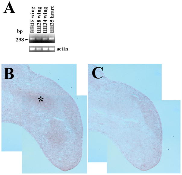
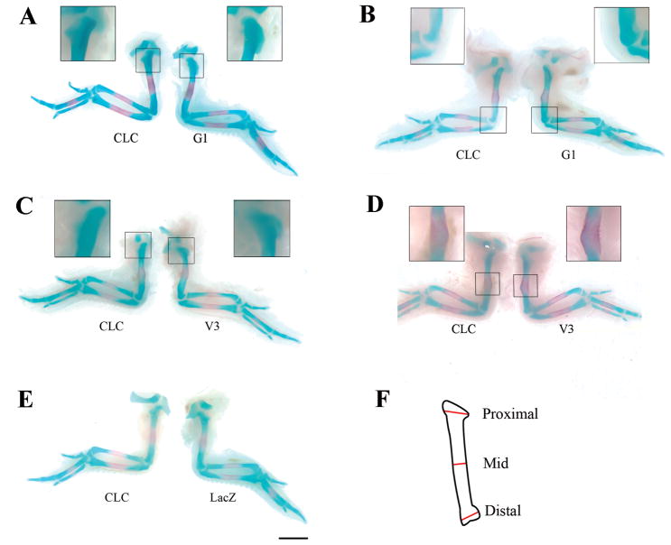
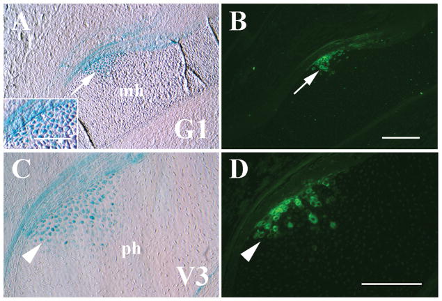
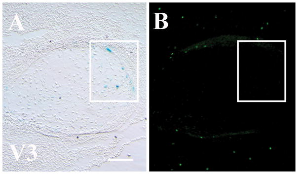
Previous work has shown that versican proteoglycan is highly expressed in the extracellular matrix of precartilage limb mesenchyme. Although much of versican's role in chondrogenesis has been attributed to its glycosaminoglycan complement, N- and C-terminal G1 and G3 domains of versican have been shown to possess distinct functions when expressed ectopically. This study was undertaken to test the hypothesis that overexpression of the versican G1 domain and short V3 isoform, comprised of only G1 and G3, in the chick wing in ovo would result in increased chondrogenesis, suggesting function for discrete versican domains in limb skeletal development. Recombinant adenoviruses encoding G1 and V3 proteins were microinjected into proximal HH19-25 chick wing buds which resulted in significant enlargement of humeral primordia at HH35. Enhanced cartilage deposition appeared due to increased chondrogenic aggregation as a result of recombinant G1 or V3 overexpression, further implicating versican in early stages of limb development.
Copyright © 2010 Wiley-Liss, Inc.
Figures
Similar articles
-
Versican knockdown reduces interzone area during early stages of chick synovial joint development.Anat Rec (Hoboken). 2012 Mar;295(3):397-409. doi: 10.1002/ar.21542. Epub 2011 Dec 20. Anat Rec (Hoboken). 2012. PMID: 22190409 Free PMC article.
-
Versican knock-down compromises chondrogenesis in the embryonic chick limb.Anat Rec (Hoboken). 2008 Jan;291(1):19-27. doi: 10.1002/ar.20627. Anat Rec (Hoboken). 2008. PMID: 18085607
-
Versican expression during synovial joint morphogenesis.Int J Biol Sci. 2007 Sep 7;3(6):380-4. doi: 10.7150/ijbs.3.380. Int J Biol Sci. 2007. PMID: 17848983 Free PMC article.
-
Versican: A Dynamic Regulator of the Extracellular Matrix.J Histochem Cytochem. 2020 Nov;68(11):763-775. doi: 10.1369/0022155420953922. Epub 2020 Sep 10. J Histochem Cytochem. 2020. PMID: 33131383 Free PMC article. Review.
-
V3: an enigmatic isoform of the proteoglycan versican.Am J Physiol Cell Physiol. 2023 Aug 1;325(2):C519-C537. doi: 10.1152/ajpcell.00059.2023. Epub 2023 Jul 3. Am J Physiol Cell Physiol. 2023. PMID: 37399500 Free PMC article. Review.
Cited by
-
Versican knockdown reduces interzone area during early stages of chick synovial joint development.Anat Rec (Hoboken). 2012 Mar;295(3):397-409. doi: 10.1002/ar.21542. Epub 2011 Dec 20. Anat Rec (Hoboken). 2012. PMID: 22190409 Free PMC article.
-
Versican G1 domain enhances adenoviral-mediated transgene expression and can be modulated by inhibitors of the Janus kinase (JAK)/STAT and Src family kinase pathways.J Biol Chem. 2017 Sep 1;292(35):14381-14390. doi: 10.1074/jbc.M116.773549. Epub 2017 Jul 6. J Biol Chem. 2017. PMID: 28684419 Free PMC article.
-
Extracellular processing of the cartilage proteoglycan aggregate and its effect on CD44-mediated internalization of hyaluronan.J Biol Chem. 2015 Apr 10;290(15):9555-70. doi: 10.1074/jbc.M115.643171. Epub 2015 Mar 2. J Biol Chem. 2015. PMID: 25733665 Free PMC article.
-
Targeted In Situ Biosynthetic Transcriptional Activation in Native Surface-Level Human Articular Chondrocytes during Lesion Stabilization.Cartilage. 2012 Apr;3(2):141-55. doi: 10.1177/1947603511426881. Cartilage. 2012. PMID: 26069627 Free PMC article.
-
Defining the earliest transcriptional steps of chondrogenic progenitor specification during the formation of the digits in the embryonic limb.PLoS One. 2011;6(9):e24546. doi: 10.1371/journal.pone.0024546. Epub 2011 Sep 13. PLoS One. 2011. PMID: 21931747 Free PMC article.
References
-
- Ang LC, Cyn MD, Zhang, Yaou MD, Cao, Liu MD, Yang BL, Young B, Kiani C, Lee V, Allan K, Yang BB. Versican enhances locomotion of astrocytoma cells and reduces cell adhesion through its G1 domain. J Neuropath Exp Neurology. 1999;58:597–605. - PubMed
-
- Aulthouse AL, Solursh M. The detection of a precartilage, blastema-specific marker. Dev Biol. 1987;120:377–384. - PubMed
-
- Barna M, Niswander L. Visualization of cartilage formation: Insight into cellular properties of skeletal progenitors and chondrodysplasia syndromes. Develop Cell. 2007;12:931–941. - PubMed
-
- Bellairs R, Osmond M. The Atlas of Chick Development. Academic Press; San Diego: 1998.
Publication types
MeSH terms
Substances
Grants and funding
LinkOut - more resources
Full Text Sources
Other Literature Sources
