. 2010 Nov;293(11):1805-15.
doi: 10.1002/ar.21216.

A novel terminal web-like structure in cortical lens fibers: architecture and functional assessment

Affiliations

A novel terminal web-like structure in cortical lens fibers: architecture and functional assessment

Kristin J Al-Ghoul et al. Anat Rec (Hoboken). 2010 Nov.

Abstract

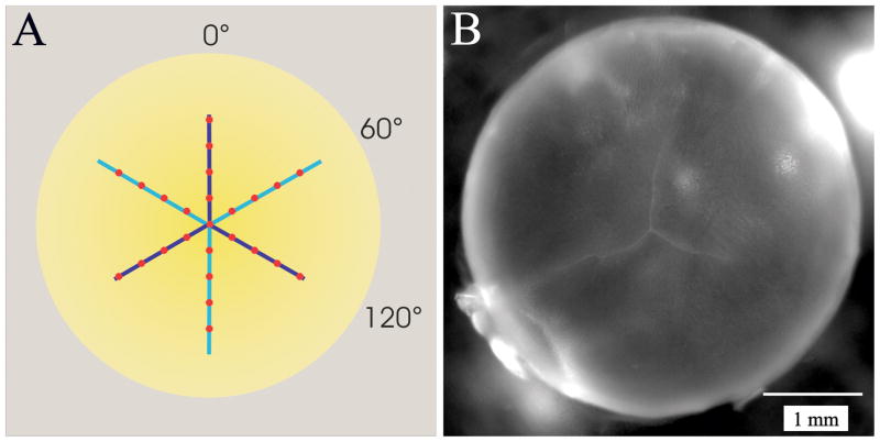
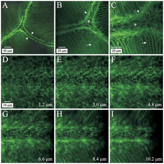
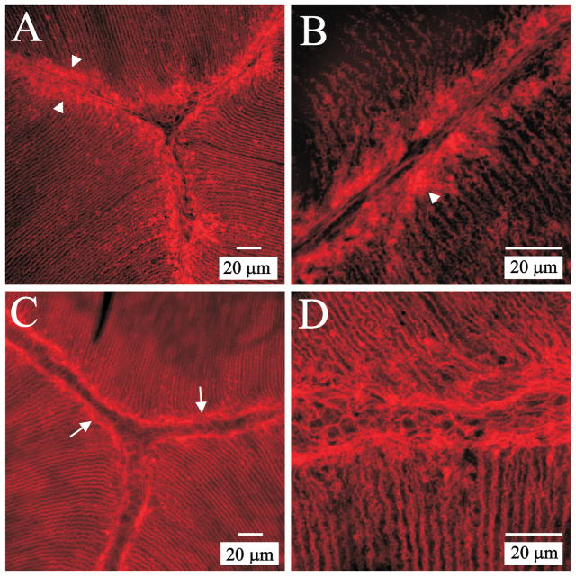
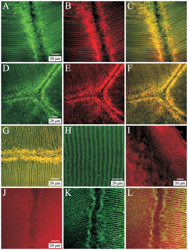
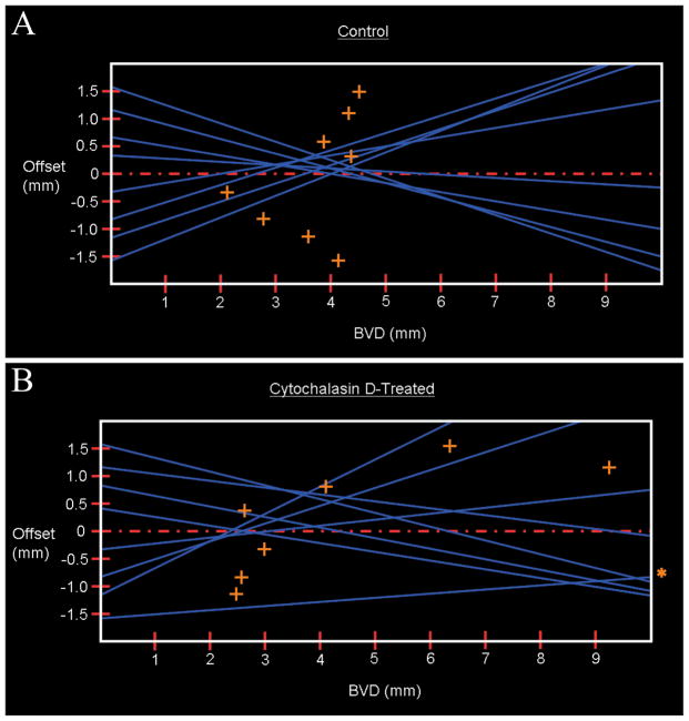
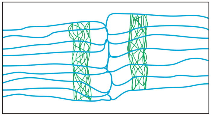
This study describes a novel cytoskeletal array in fiber cells of the ocular lens of the rat and shows its relationship to the classical terminal web of other epithelial tissues. Naive adult Sprague-Dawley rats (n = 28) were utilized. F-actin, fodrin, myosin IIA, and CP49 distribution was assessed in anterior and posterior polar sections. For functional analysis, lenses were cultured with or without cytochalasin-D for 3 hr, then processed for confocal microscopy or assessed by laser scan analysis along sutures. Phalloidin labeling demonstrated a dense mesh of F-actin adjacent to posterior sutural domains to a subcapsular depth of 400 μm. Anterior polar sections revealed a comparable actin structure adjacent to anterior suture branches however, it was not developed in superficial fibers. Fodrin and myosin were localized within the web-like actin apparatus. The data was used to construct a model showing that the cytoskeletal array is located within the blunt, variable-width fiber ends that abut at sutures such that the "terminal web" flanks the suture on either side. Treatment with cytochalasin-D resulted in partial disassembly of the "terminal web" and perturbed cellular organization. Laser scan analysis revealed that cytochalasin-D treated lenses had significantly greater focal variability than control lenses (P = 0.020). We conclude that cortical fibers of rat lenses contain a bipolar structure that is structurally and compositionally analogous to classical terminal webs. The results indicate that the lens "terminal web" functions to stabilize lens fiber ends at sutures thus minimizing structural disorder, which in turn, promotes the establishment and maintenance of lens transparency.


Figures

Figure 1
Figure 1
A. Diagram demonstrating the position of laser beam penetrations with respect to the lens sutures. Anterior suture branches are in light blue; posterior suture branches are in dark blue (seen through the lens). Each series of scans passes along an anterior and a posterior suture branch. B. Photograph of the posterior aspect of a rat lens showing the typical inverted Y suture pattern.
Figure 2
Figure 2
LSCM images of phalloidin-FITC labeled posterior polar lens sections. A. Low magnification of the confluence of the posterior sutures approximately 10 μm deep to the CFI. The typical inverted-Y suture pattern is apparent. B. Medium magnification of posterior sutures at about 50 μm from the CFI. The sutural region is flanked by the web-like mesh, which appears to attach to the actin filaments in both the fiber ends and the lateral fiber borders. C. High magnification of a posterior suture branch approximately 275 μm deep to the CFI. D–I. Non-adjacent optical sections from a z-series taken near the proximal end of a posterior suture branch, showing the formation of the web-like F-actin structure. Distance from the CFI is indicated in the lower right corner of each panel; D–I are at identical magnification. By approximately 3.5 μm from the CFI an indistinct mesh of F-actin was present flanking the forming suture branch. At 10 μm deep to the CFI, the F-actin underlying lateral fiber membranes is contiguous with the well-formed web-like structure.
Figure 3
Figure 3
LSCM images of phalloidin-TRITC labeled anterior polar lens sections. A. Low magnification of the confluence of anterior sutures approximately 10 μm deep to the EFI shows accumulations of F-actin at fiber ends. B. Higher magnification of an anterior suture branch in another lens (approximately 15 μm deep to the EFI) reveals that F-actin is not organized into a mesh-like web in anterior ends of superficial maturing fibers. C. Low magnification of the confluence of anterior suture branches about 60 μm from to the EFI shows a prominent ‘terminal web.’ D. Higher magnification of an anterior suture branch in another lens approximately 75 μm deep to the EFI shows the F-actin web architecture is comparable to that at posterior ends of fibers.
Figure 4
Figure 4
LSCM localization of terminal web proteins within the F-actin web of lens fibers. A–C. Double labeling for myosin (A) and F-actin (B) at a posterior suture branch. The merged image (C) shows that myosin and actin were largely colocalized within the web-like mesh as well as along lateral aspects of fiber segments. Myosin was present in fiber ends as a diffuse plaque (consistent with previously published data (Lu et al., 2008). A–C are at identical magnification. D–F. Low magnification image series of double labeling for fodrin (D) and F-actin (E) in a posterior polar section. The merged image (F) demonstrates a high degree of colocalization. D–F are at the same magnification. G. Merged higher magnification view of a posterior suture branch double labeled for fodrin and F-actin shows the extensive co-localization of these components within the ‘terminal web’ as well as along lateral fiber membranes. H. Positive control for fodrin specificity. Fodrin localization in equatorial segments of cortical fibers revealed that labeling was most prominent along the short sides of hexagonal fiber profiles. I. Positive control for CP49 specificity. Equatorial segments of cortical fibers demonstrated peripheral localization of CP49 in superficial fibers and a more diffuse, cytoplasmic labeling in deeper cortical fibers. J–L. Double labeling for CP49 (J) and F-actin (K) at a posterior suture branch revealed the diffuse pattern of CP49 label both within the ‘terminal web’ and along posterior segments of fibers. The merged image (L) emphasizes the lack of strong CP49 labeling within the F-actin web adjacent to the posterior suture branch. J–L are at identical magnification.
Figure 5
Figure 5
LSCM visualization of cytochalasin D-treated (A and C) and control (B and D) lenses. Panels A and B show posterior sutures at approximately 20 μm and 40 μm from the CFI, respectively; panels C and D illustrate anterior sutures at about 110 μm and 85 μm from the EFI, respectively. Specimens were double labeled for F-actin (green) and wheat germ agglutinin (red) in order to localize the actin cytoskeleton and outline the fiber membranes. Cytochalasin D treatment resulted in partial disassembly of the ‘terminal web’ resulting in disarray of fiber end arrangement at both the posterior (A) and anterior (C) sutures. In contrast, fiber end segments outside of the sutural domains were intact and appeared undisrupted by cytochalasin D treatment (arrows).
Figure 6
Figure 6
Representative laser scan profiles of OS lenses (A) which were untreated controls and OD lenses (B) which were treated with Cytochalasin D. The more diffuse pattern of laser scans for the cytochalasin-D treated lenses illustrates their greater variability in BVD. The offset distance is the distance from the lens center for each scan series. Blue lines represent individual laser beam penetrations; orange crosses denote the BVD for each laser penetration. A. Mean BVD = 3.6 mm. B. Mean BVD = 6.3 mm. Laser penetration at offset −1.5 (indicated by asterisk) had a BVD of 20.3 mm.
Figure 7
Figure 7
Two-dimensional model of murine fiber end segment architecture showing the relative position of the ‘terminal web’ within a single growth shell. End segments of fibers (membranes shown in blue) are non-uniform in width and have blunt ends where they abut with opposing fiber ends to form a suture. The F-actin of the web-like cytoskeletal apparatus (shown in green) is located within fiber ends such that the ‘terminal web’ flanks the suture on either side. Neither the undulating surface topology of lateral fiber membranes nor the cortical actin beneath them is depicted.

Similar articles

Cited by

References

    1. Al Awqati Q, Vijayakumar S, Takito J, Hikita C, Yan L, Wiederholt T. Terminal differentiation in epithelia: the Hensin pathway in intercalated cells. Semin Nephrol. 1999;19:415–420. - PubMed
    1. Al Awqati Q, Vijayakumar S, Takito J, Hikita C, Yan L, Wiederholt T. Phenotypic plasticity and terminal differentiation of the intercalated cell: the hensin pathway. Exp Nephrol. 2000;8:66–71. - PubMed
    1. Al-Ghoul KJ, Kuszak JR, Lu JY, Owens MJ. Morphology and organization of posterior fiber ends during migration. Mol Vis. 2003;9:119–128. - PubMed
    1. Al-Ghoul KJ, Novak LA, Kuszak JR. The structure of posterior subcapsular cataracts (PSCs) in the Royal College of Surgeons (RCS) rats. Exper Eye Res. 1998;67:163–177. - PubMed
    1. Alberts B, Johnson A, Lewis J, Raff MC, Roberts K, Walter P. Molecular biology of the cell. 4. New York: Garland Science; 2002.

Publication types

MeSH terms

LinkOut - more resources