

doi: 10.1186/1758-2555-2-21.
Medial and lateral discoid menisci: a case report
Affiliations
- PMID: 20731824
- PMCID: PMC2936413
- DOI: 10.1186/1758-2555-2-21
Item in Clipboard
Medial and lateral discoid menisci: a case report
Sports Med Arthrosc Rehabil Ther Technol.
.
Abstract
Discoid menisci on both medial and lateral tibial plateau are very rare abnormalities. We report a 44-year-old woman with bilateral medial and lateral discoid menisci. She also had anomalous insertion of discoid medial meniscus to anterior cruciate ligament, and pathologic medial patellar plica on the right knee. Meniscectomies has been performed for her torn discoid menisci with satisfactory result on the latest follow-up.
Figures

Anteroposterior and lateral standing radiographs of the both knee. (A) Anteroposterior and (B) lateral (right and left) radiographs showing bilateral widening of lateral joint spaces, and increased concavity and subchondral sclerotic of the medial tibial plateau.

MRI image of the right knee (A) T1-weighted sagittal MRI of the right knee showing medial discoid meniscus with a horizontal tear (indicated by red arrow) and (B) showing lateral discoid meniscus; whereas (C) the T2-weighted coronal MRI showing medial discoid meniscus with tear (indicated by red arrow) and lateral discoid meniscus with no tear.
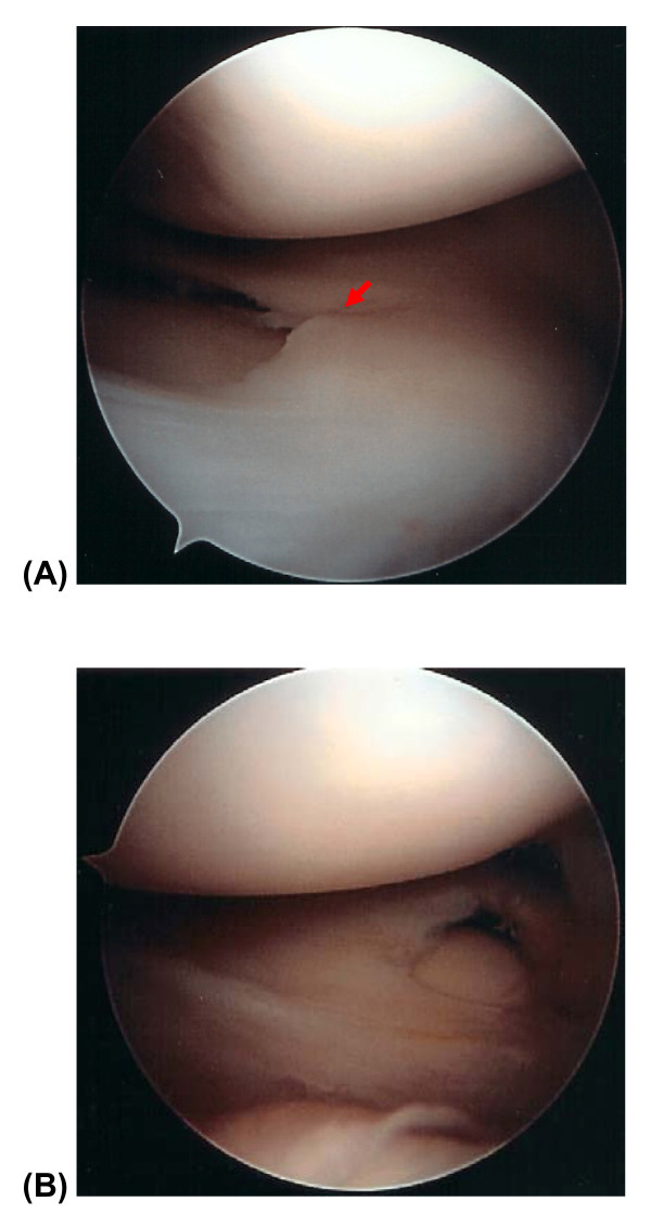
Arthroscopic view of the right knee. Arthroscopic examination of the right knee showed (A) an incomplete medial discoid meniscus with horizontal cleavage tear (indicated by red arrow), and (B) a complete lateral discoid meniscus without tear.

Original scan operating report done by senior author. Operating scheme describing incomplete left medial discoid meniscus with flap tear and incomplete left lateral discoid meniscus with horizontal tear. (A) Before meniscectomy. (B) After subtotal meniscectomy of the discoid medial meniscus and reshaping of the discoid lateral meniscus.
References
-
- Young RB, Cleland J, MacKay JY. Memoirs and memoranda in anatomy. Vol. 1. London, Williams and Norgate; 1979. The external semi-lunar cartilage as a complete disc; pp. 179–80.
-
- Cave EF, Staples OS. Congenital discoid meniscus: A cause of internal impingement of the knee. Am J Surg. 1941;54:371–376. doi: 10.1016/S0002-9610(41)90383-5. - DOI
-
- Jeannopoulus CL. Observations on the discoid menisci. J Bone Joint Surg Am. 1950;32:649–652. - PubMed
-
- Murdoch G. Congenital discoid medial semi-lunar cartilage. J Bone Joint Surg Br. 1956;38:564–566. - PubMed
LinkOut - more resources
Full Text Sources
